Overview

Fast-Forward to Next Generation Imaging

The Aperio GT 450 DX is designed to scale up digital pathology operations by increasing throughput in high volume clinical pathology laboratories. The system uses innovative color calibration technology to output consistent high-quality images enabling on-time, on-screen diagnosis. Aperio GT 450 DX is an automated, high capacity digital pathology slide scanner that offers exceptional image quality, rapid delivery of cases and high-performance streamlined digital viewing built with the pathologist in mind.

  • Improve case turnaround with no touch scanning and continuous loading of the racks directly from the HistoCore SPECTRA Workstation (Stainer & Coverslipper)
  • Increase IT security and control with a dedicated Scanner Administrator Manager DX (SAM) server and software that allows you to set up and monitor multiple Aperio GT 450 DX scanners at a time. No more single workstations for each scanner
  • Ensure excellent image quality with Leica optics
Contact Us to get a quote. Call 00 308 321 0320 to speak with a representative.

For In Vitro Diagnostic Use.


Aperio GT 450 DX DOWNLOAD THE BROCHURE NOW!


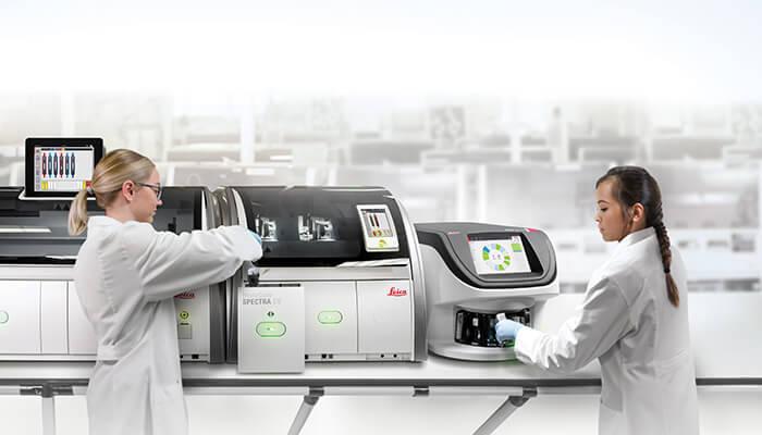
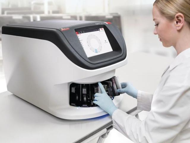
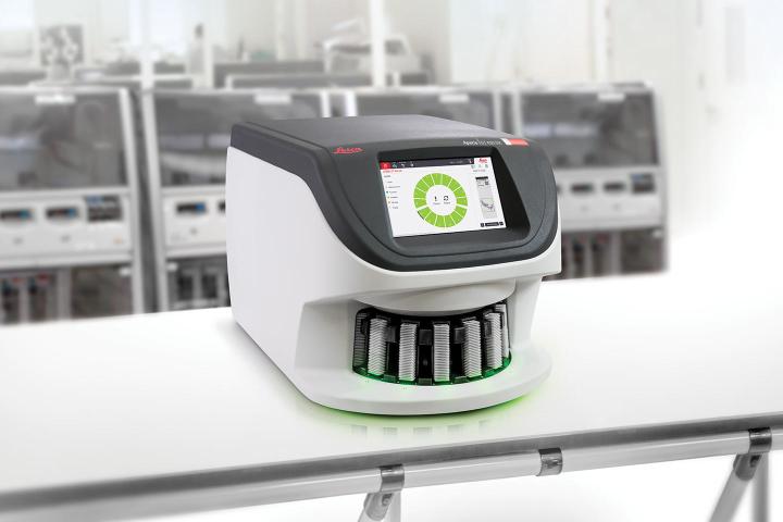
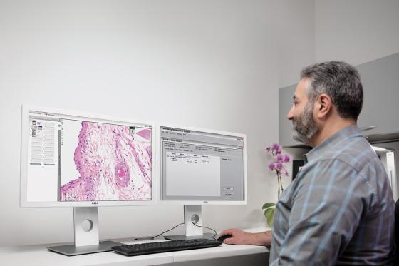
Fast and Easy to Operate for Histotechnicians

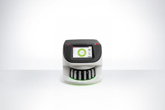
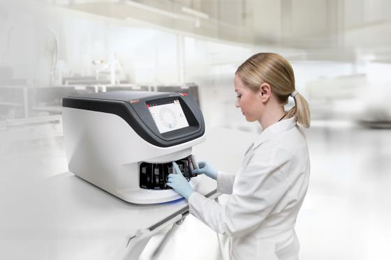
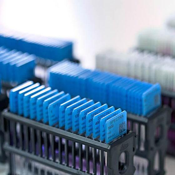
The Aperio GT 450 DX is simple to operate and helps histotechnicians deliver rapid results with confidence. Complete scanning quickly and easily so you can focus on other tasks in the lab: 15 racks of 30 slides (450 slides total) can be loaded directly from HistoCore SPECTRA CV Coverslipper into the GT 450 DX. The slides are then automatically scanned at 81 slides per hour at 40x for a 15mm x 15mm area.

  • No touch continuous loading during scanning
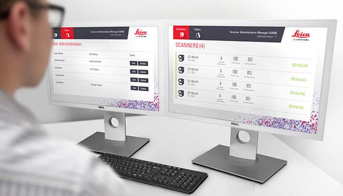
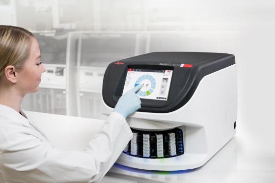
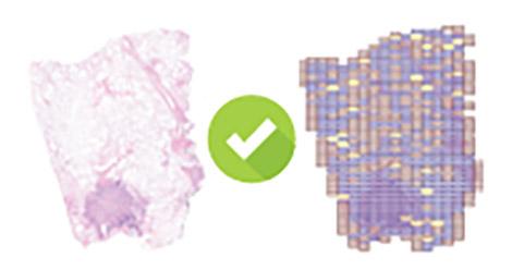
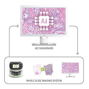
  • Automated IQ checks during each scan to ensure quality
  • Assign priority cases
  • Every slide is calibrated with each scan
  • 450-slide capacity packed into a small size fits on the lab bench
  • 99.5% accurate tissue finder that allows you to adjust if needed*
  • 100% first-time barcode scan success rate*

*VP-0463 validation results

From the lab to the IT room to the office, the Aperio GT 450 DX was designed to scale up digital pathology operations for histotechnicians, IT professionals and pathologists.

Image
Fast and Easy to Operate
Aperio GT 450 DX can be loaded directly from HistoCore SEPCTRA CV Coverslipper

Secure, Scalable Architecture for IT Professionals

The Aperio GT 450 DX offers fast, secure, and flexible IT architecture. A centralised SAM server and dedicated software provides you with a data management solution that can remotely set up and monitor multiple Aperio GT 450 DX scanners at a time.*

  • Comprehensive cybersecurity including encryption and login controls
  • centralised scanner management to set up and monitor multiple scanners
  • Two 10GB network connections on the SAM server
  • Scalable set up for hub and spoke configurations
  • Supports dedicated logins for each Aperio GT 450 DX scanner

*See Resources > FAQ for more info on SAM

Image
Secure, Scalable IT Architecture

Excellent Image Quality for Pathologists

The Aperio GT 450 DX uses a high=performance objective manufactured by Leica Microsystems, renowned for producing world-class optics since 1849. This historic craftmanship is now built into the today’s Aperio GT 450 DX, which is designed to rapidly deliver images with exceptional quality for Pathologists.

  • Leica optics deliver exceptional image quality
  • Every slide is calibrated during scanning to ensure the best quality image
  • Automated image quality assessment, so you don’t have to send slides back for re-scans
  • 99.5% accurate tissue finder finds faint tissue while excluding pen marks, dust, and residue
Image
Excellent Image Quality with Custom-Made Leica Objective
Custom-made Leica objective for next generation scanning,
Image
csm_GT_450_Image_for_website_a20f3ca405

Image Quality Tested by Pathologists for Pathologists

(for diagnostic review, use Aperio DX Viewer on the Aperio DX Viewing Station, which includes a calibrated monitor)

Image
Mitotic Events (Breast)
Image
Intercellular Bridges
Image
Chromatin
Chromatin
Image
Nucleoli
Nucleoli

Precision, Potential to Scale, and Peace of Mind

Digital pathology scan speeds have not always been fast enough at 40x magnification to keep up with high-volume scanning (120k+ slides per year). Real-Time Focusing (RTF)** offers a potential solution to this problem. It’s a novel method to capture a digital image that combines an imaging line sensor and a focusing line sensor.

  1. While scanning, the focusing line sensor receives focusing data of the tissue.
  2. Proprietary algorithms determine the best focus value.
  3. Then, each best focus value is treated like a control loop with the mechanics, feeding the best focus value to the objective height on the fly.
  4. The positioning adjustments of the objective are performed continuously (in real time) in order to enable the imaging line sensor to capture the image at its best possible focus. Hence, real time focusing.

**US Patent no. 9,841,590

Image
Cellular Groves (Thyroid)

Excellent Image Quality with Custom-Made Leica Objective

The high-performance objectives included with the Aperio GT 450 DX is specifically designed to maximise field of view for high speed digital pathology scanning. Most objectives are designed similarly to the human eye: round, which focuses light in the center and limits field of view. The objectives on the Aperio GT 450 DX includes an extra-wide flat field correction that enables a much larger field of view (1mm) that can accommodate extremely large digital images for fast scans and 0.26 um/pixel resolution at 40x magnification.

Image
Excellent Image Quality with Custom-Made Leica Objective

Specifications

Imaging, Capacity & Operations
Dimensions & Weight
IT Requirements & Integration
Security Features
Slides, Coverslips & Labels

Downloads

View all available downloads below

Similar Products

HistoCore SPECTRA Workstation

HistoCore SPECTRA Automated Slide Staining Workstation

The highly automated workstation solution to manage increasing workloads and high throughput demands. Specifically designed to deliver timely and consistent results from slide 1 – 1600.

Knowledge Hub

From tissue processing to Digital Pathology, our solutions empower labs with efficiency, precision, and reliability.

top consideration when buying a scanner
Top Considerations When Buying a Digital Pathology Scanner

Are you thinking about purchasing a digital pathology scanner? We offer some tips for things to consider during the buying process.

4-tips-for-digital-pathology-slide-scanning-640x480
Four Tips for Digital Pathology Slide Scanning

Do you want to get better digital images from your glass slides? Here are some tips to help you get the best out of your digital pathology line scanner: