
Navigator through the Brain - Stereotaxic Atlases for Neuroscience Research
We understand you are working with Elsevier on the release of a new automated atlas. Can you describe it?
The automated onscreen atlas now contains 3Dmaps of rat and mice brain, and it will contain human brain. Next, we will add monkey and possibly other species. The atlas can be rotated by any angle of approach and then sectioned by a software-free cutting tool at any desired angle. It is like the sectioning of a brain at any angle, not necessarily in the three cardinal planes.
For rat and mouse stereotaxic surgery, it is helpful for interpreting brain sections that are not cut in the plane of the atlas; that is, are laterally asymmetrical, or, more frequently, not cut in the skull-flat position top to bottom. For example, the dorsal part of the brain may be some sections more rostral or caudal than the ventral part of the brain. To interpret one section on the microscope, you would have to go through five pages of an atlas, which is cumbersome and can lead to errors. But if you can resection the atlas data to produce a cut that better resembles what is seen under the microscope, it is very helpful. Even in my own lab, a brain is sometimes sectioned, so the coronal sections are not perpendicular to skull-flat and students ask, “What do I have here?” I find that the section is somewhere between the horizontal and coronal plane.
It is hard to figure out where a given point is, if you are not in the atlas plane of section.
If you take a horizontal line, and you are at the fasiculus retroflexus and the medial lateral plane is symmetrical, then you have the same structure on the other side. You cannot be anywhere else. But if you go higher to the habenula or down below to the hypothalamus, you don’t know. You have to find a landmark at the same horizontal level. Often the brain does not have landmarks that you can be absolutely certain about.
Are you planning future atlas projects?
In clinical psychology they call it ‘perseveration.’ [laughter] You just can’t quit! Although we haven’t sectioned it yet, we are working on an atlas of the marmoset brain. The marmoset is the size of a small rat, yet it is a primate. The work we have done, so far in the cortex of the marmoset indicates that its cortical areas of cerebral cortex are the same as the cortical areas of human brain.
We want to do an atlas of the human cortex, which could be an atlas of the entire human brain. There hasn’t been a good atlas of the human cortex since the work of Von Economo in the 1920s. Now it is Brodmann who is followed, but Von Economo is better. But even Von Economo does not have the accuracy we find in the monkey atlases, which are far better at delineating cortex. The new monkey atlas that we have just done is the most accurate atlas of a primate ever produced. The cortical delineations are state-of-the-art.
What inspired you to go into atlas work?
I went on a sabbatical to Cambridge 1976, where my work involved cutting brain in horizontal sections to best interpret my knife cuts. If I cut coronal, I could not see what I was doing, I only saw cut lines. But I needed to see pileup accumulation of immunoreactive material in one direction and depletion in the other direction. There was no atlas of rat brain that showed horizontal sections, so I thought of building and publishing one. The paper did well but what people really wanted was coronal. Although what we had published was better for stereotaxic and had better delineations than König and Klippel, coronal was clearly what we had to do.
Did you use fresh tissue (to avoid shrinkage) on that early atlas?
Yes. I had picked up the technique of staining for acetylcholinesterase, and I thought I could see the brain better. There were no ambiguities in many instances, so I had the idea that we could do a better atlas. Horizontal was of course easier, but the game is played in another field, coronal. Although it was very difficult, we did the coronal sections over three years. I was inserting 15 marker electrodes from rostral to caudal every 2 mm plus three horizontal electrodes in each brain, plus two sagittal electrodes to mark the coordinate system.
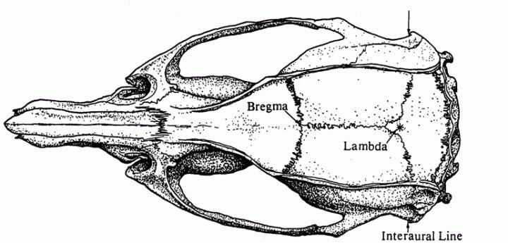
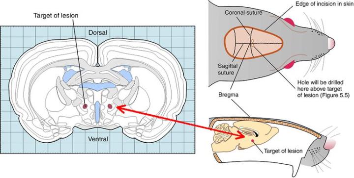
By the way, at that time we defined the now standard reference position for the skull, the skull-flat position, with Bregma and Lambda at the same vertical position; and how to locate them not necessarily exactly where the sutures cross, but where the lines of best fit would cross the midline suture.
Wasn’t Bregma being used in the 1980s?
Yes, as the zero point, but at some elevation of the incisor bar, rather than skull-flat. We defined and implemented the skull-flat position for our atlases [and it improved reproducibility].
As we were taking the brain out, it would assume the shape of whatever surface it was on. I was trying to get all my electrodes to come in one section; an aligned cut of brain. I then had the idea to freeze the whole head together with the brain inside. I prized the frozen bone off the frozen brain, which didn’t give the brain a chance to assume the shape of what it was placed on. At that point, I really made progress.
It must have been a really difficult dissection
Yes, but it was really the issue of getting accurate coordinates. It was one year’s work of starting at 9 a.m. and finishing, if the brain was promising, at 4 a.m. the next morning. I worked like this for a year to ensure that each of the cuts was exactly right. I was always getting so close.
From the olfactory bulb to the spinal cord!
All in three cardinal planes. But I was convinced that the König and Klippel atlas was not really helpful to scientists, as it omitted many parts of the brain and the point of Bregma, the most frequently used reference point in stereotaxic surgery. It was not useful for stereotaxic surgery.
In addition, all the coordinates were inaccurate, as the brain had shrunk
Yes, it was not only shrunk, but the atlas described 150-gram female rats whereas most researchers use 300-gram males. There is a big difference between these brains. But my greatest difficulty with it was in my graduate years. I was to be instructed by my professor on how to do stereotaxic surgery on a rat, but the rat declined to go under anaesthetic. The professor said, “Look, I have run out of time, but when he goes under, implant the electrode in the hypothalamus. Find the coordinates from the atlas.” In my rush to operate on the animal before he woke up, I failed to read the introduction where it was clearly stated that the stereotaxic zero point of the atlas coordinates was not the zero point of the stereotaxic instrument, but 4.9 mm above the earbar plane. In targeting the hypothalamus, I missed the brain by 4.9 mm. And I thought that any psychologist, as I was then, could do better than that. The whole point is to have an atlas to depend on, not just refer to. At that time, you always had to derive your coordinates experimentally.
As a side story, when I tried to publish this new atlas, Elsevier replied that one of their advisors told them not to publish it, and there was a rhetorical question, “Why yet another atlas of the rat brain?” We knew the need of another atlas, as the König and Klippel atlas did not display the pons, the medulla, the cerebellum, the cortex, the spinal cord, and the point of Bregma. It was not useful for stereotaxic surgery, it was not there. But eventually Academic Press, now owned by Elsevier, published it.
What advice would you give to young neuroscientists?
What I have found in neuroscience, is that if you have the hardware, don’t let a project wait. Work straight through the weekends. Perseverance, not [necessarily] perseveration, but perseverance. There is more perspiration than inspiration in science. I have seen very clever students fall by the wayside, and others succeed because they were more determined to succeed. My advice is, don’t wait for inspiration in the first sentence of your paper. Just write something and keep going. More specifically to neuroscientists, get to know the concept of neuromeres.
If you look in the Atlas of the Rat Brain in Stereotaxic Coordinates, Compact 6th Edition, you will see an introduction to the concept. There are 15 diagrams where we explain it for the reader who is new to the idea. This concept will have a big impact on our understanding of neuroanatomy and function.
The idea of neuromeres?
Yes, a neuromere is a developmental unit. The same gene groups are responsible for patterning in the nervous system of the human and the mouse. They are also involved in the segmenting the body of Drosophila. These genes act throughout evolution. We are now just appreciating the importance of them in the organisation of the brain stem, how it is put together and so how it will function.
Related Content


Leica Biosystems Knowledge Pathway content is subject to the Leica Biosystems website terms of use, available at: Legal Notice. The content, including webinars, training presentations and related materials is intended to provide general information regarding particular subjects of interest to health care professionals and is not intended to be, and should not be construed as, medical, regulatory or legal advice. The views and opinions expressed in any third-party content reflect the personal views and opinions of the speaker(s)/author(s) and do not necessarily represent or reflect the views or opinions of Leica Biosystems, its employees or agents. Any links contained in the content which provides access to third party resources or content is provided for convenience only.
For the use of any product, the applicable product documentation, including information guides, inserts and operation manuals should be consulted.
Copyright © 2026 Leica Biosystems division of Leica Microsystems, Inc. and its Leica Biosystems affiliates. All rights reserved. LEICA and the Leica Logo are registered trademarks of Leica Microsystems IR GmbH.