
概要

Product SKU: Workstation Kit - 14051254355, ST - 14051254354, CV - 14051454200
Flexibility with Consistent Quality from Slide 1 to 1,600

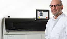
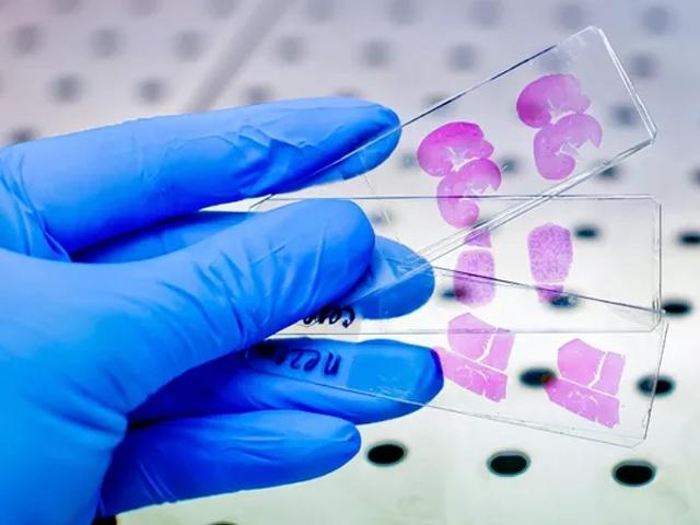
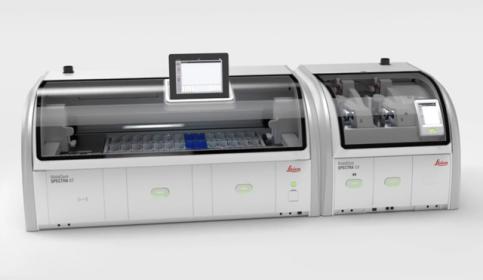
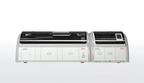
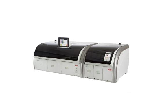
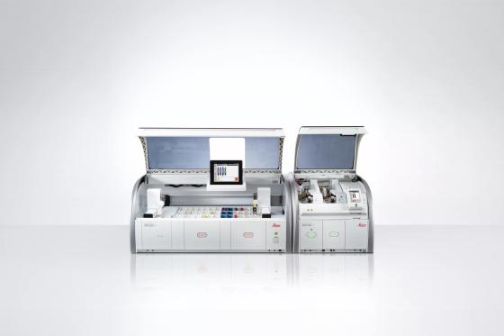
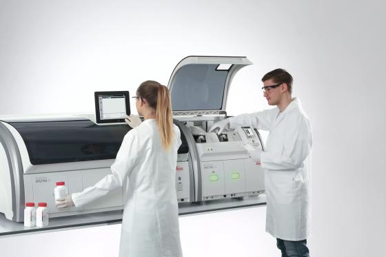
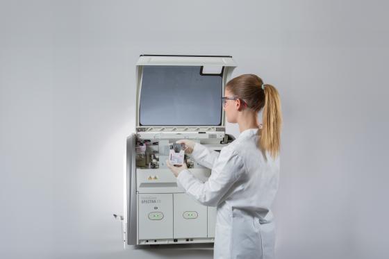
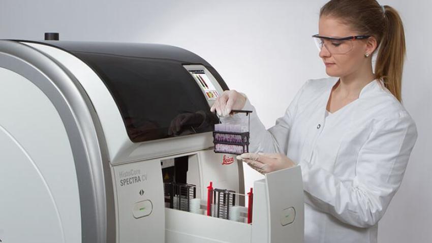
Designed to adapt to your lab’s evolving needs, the HistoCore SPECTRA Workstation combines exceptional workflow flexibility with high-quality, consistent staining results. Whether processing a few slides or larger workloads, HistoCore SPECTRA WS enables labs to run H&E and special stains simultaneously while supporting reproducibility, operational efficiency, and confidence in every slide.

  • Flexible throughput: Seamlessly manage varying workloads, with the capacity to process up to 360 slides per hour while maintaining consistent, high-quality staining results.
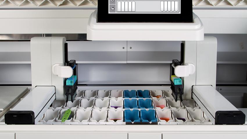
  • Parallel processing versatility: Independently run H&E and special stains at the same time across 52 stations, providing flexibility while preserving reproducibility.
  • Quality-driven consistency: Dedicated consumables help ensure consistent staining outcomes while minimizing variability and reducing the need for manual corrections.
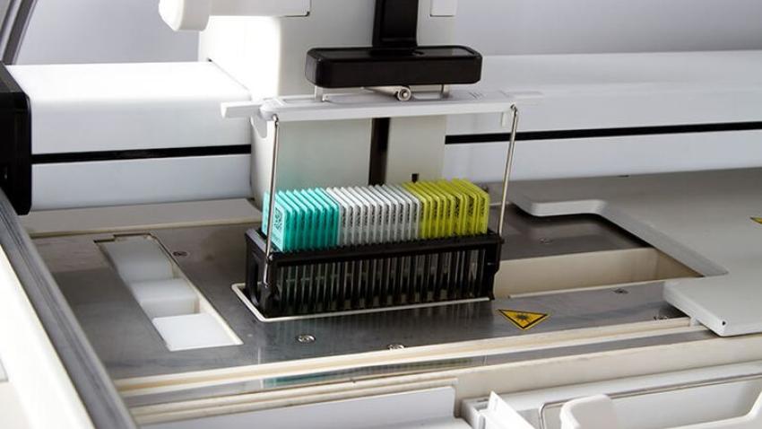
  • Reliable, automated tracking: Integrated barcode reading enhances traceability and workflow efficiency, helping maintain accuracy and quality control across every slide.

With a 5-minute coverslipping drying time, slides are ready to be scanned—supporting both rapid turnaround and uncompromised quality at every step.

購入に関するご相談 ご購入前のお問い合わせ。

For in vitro diagnostic use.


NOW AVAILABLE HistoCore LIGHTNING C Laser Cassette Printer


特徴

Automated Routine & Special Staining with Dual Robotic Arms
Smart Reagent Management for Cost and Quality Control
Confident Workflow with Automated Slide Tracking
Error Reduction with Intelligent Protocol Automation
Unmatched Throughput and High-Capacity Output for Busy Labs
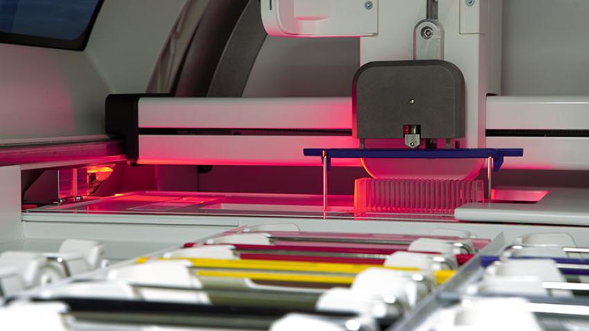
Smarter Coverslipping for Higher Efficiency
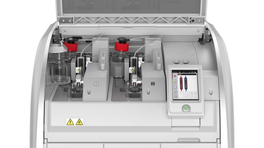
Improved Layout for a More User-Friendly Experience

Consumables

Consistent Quality You Can Trust

Reliable Results from Slide 1 to 1,600

Achieve confidence in every batch. Leica Biosystems’ validated protocols—paired with the HistoCore SPECTRA H&E Staining Systems—help ensure consistent, high-quality results, minimizing day-to-day variability and supporting faster, more accurate diagnoses.

Image
スライド 1 枚目から 1600 枚目に至るまで、一貫した品質を実現

Ready-to-Use Consumables for Superior Results

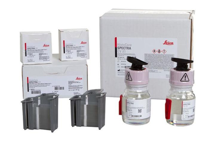
  • HistoCore SPECTRA CV X1 Mounting Media
    A rapid-drying, xylene-based formula designed for efficiency and durability. With antioxidant protection, it prevents stain fading and artifacts while allowing slides to be handled immediately after mounting. Compatible with xylene, toluene, and most substitutes.
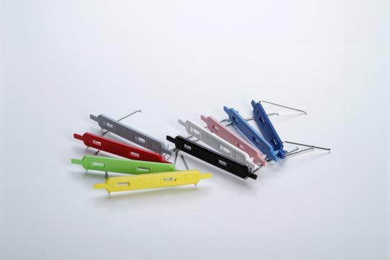
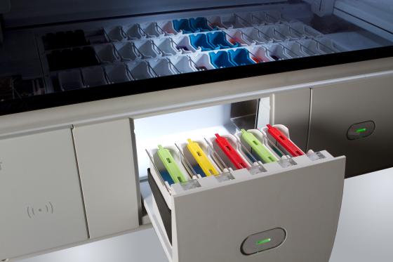
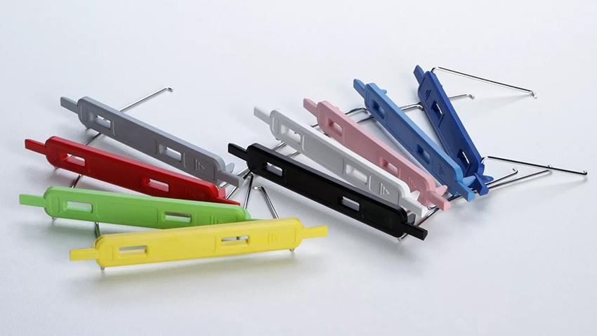
  • HistoCore SPECTRA CV Coverglass
    Delivered in ready-to-use cartridges it minimize fingerprints while improving ease of use. Enhanced packaging helps reduce breakage and protects against humidity, helping support consistent performance.

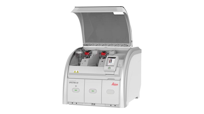
The HistoCore SPECTRA CV combines speed, precision, and intelligent design to deliver exceptional throughput, dependable quality, and an efficient, safe coverslipping workflow.

Image
Ready-to-Use Coverglass and Mounting Media

仕様

HistoCore SPECTRA ST Stainer
HistoCore SPECTRA CV Coverslipper
HistoCore SPECTRA H&E Staining Systems
HistoCore SPECTRA CV Consumables

構成

Workstation
Stainer
Coverslipper
Product configuration codes
Workstation Solution
Stainer Only
Coverslipper Only
HistoCore SPECTRA ST 全自動染色装置(14051254354)
—
HistoCore SPECTRA CV 全自動封入装置(14051454200)
—
HistoCore SPECTRA CV カバーガラス(3800152)と封入剤(3801733)
—
HistoCore SPECTRA ワークステーションキット/搬送ユニット(14051254355)
—
—

Resources

Customer Testimonials

Image
Histocore_SPECTRA_CV_Coverslipper_Reduces_Drying_Completion_Time

HistoCore SPECTRA Coverslipper reduces turnaround time by 47% from glass coverslipping to slide drying process completion.

An independent university pathology hospital compared the HistoCore SPECTRA Coverslipper with the Sakura Tissue Tek® Film Coverslipper. Result: The use of the built-in oven reduced the drying process time by 47% compared to using the drying station of their existing film coverslipper.

Read more

Image
Wisconsin_Diagnostic_Laboratories_Improves_Stain_Quality_and_Workflow

"The fact that the HistoCore SPECTRA stainer stains and maintains the same quality from the first slide to the sixteen-hundred is amazing to me.”

Wisconsin Diagnostic Laboratories Histology Department in Milwaukee, Wisconsin, handles more than 24,000 cases and 500,000 blocks every year. They needed to update their staining equipment after 20 years of service and looked for a replacement that would continue their long tradition of highly accurate, efficient service.

Read more

Image
Inform_Diagnostics_teams_up_with_Leica_Biosystems

“The nuclear detail is of excellent quality, much like looking at a picture in a book. The pathologists just love the quality of the slides.”

Always looking to improve its quality and streamline operational efforts, Inform Diagnostics teamed up with Leica Biosystems to assess whether the HistoCore SPECTRA systems provided a cost-effective update to Inform Diagnostics’ staining technology that would meet or exceed its industry-leading quality standards.

Read more

関連製品

bond-iii

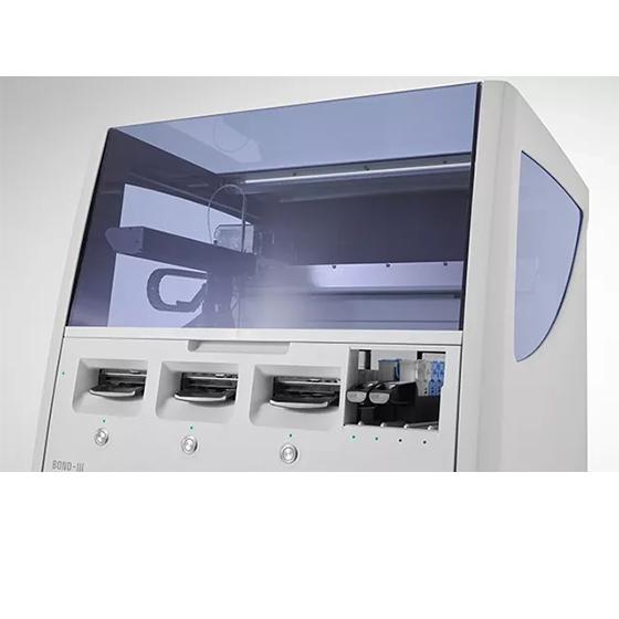
BOND-III完全自動IHCおよびISH染色システム

品質の高いIHC染色を、平均わずか2.5時間の染色時間で実施します。スピード、品質、効率性に優れた全自動免疫染色装置です。

ナレッジハブ

From tissue processing to Digital Pathology, our solutions empower labs with efficiency, precision, and reliability.

H and E Stain - A lesson on Consistency and Reproducibility by Carolyn Doan

HE染色 : 一貫性と再現性を保つための動画コンテンツ

Producing an H&E stained slide is a process. It starts way before the slide is loaded on a stainer or moved down through containers by hand. Producing quality, consistent and reproducible H&E stained slides is a process as well as a lesson in...

KP_Hero_Image_TANGERINE_01-640x480

ムチン評価のための特殊染色技術

Special stains that are used for the evaluation of mucins, mucin-like molecules and other carbohydrate containing macromolecules remain in demand and are utilized frequently in the histology laboratory.

H-E-Staining-Overview-A-Guide-to-Best-Practices-640x480

HE染色の概要:ベストプラクティスへの手引き

日常的な診断では、ヘマトキシリンとエオシン(HE)の使用が、病理医が細胞や組織構造の詳細を見るために断然好まれています。

KP_Hero_Image_MERLOT_01-640x480

一般染色および特殊染色の手引き

HE染色(ルーチン染色)および特殊染色は顕微鏡下で元来透明な組織標本を視覚化するために必要な、組織病理学の診断に不可欠なプロセスです。ここではそのHE染色や特殊染色の基本的なワークフローを各ステップごとに解説します。