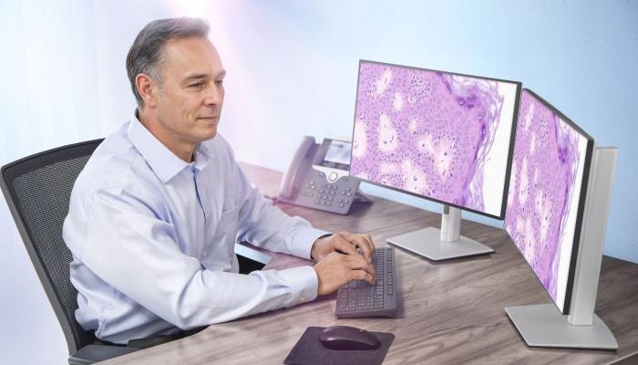
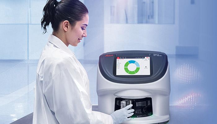
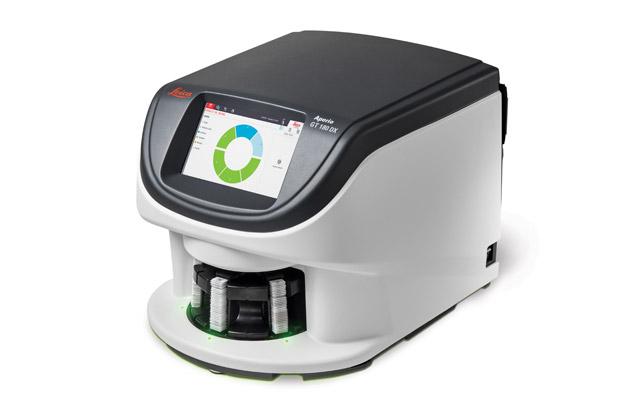
Overview

It's Time for a 180 - Because Proven Technology Means Better Pathology

The Aperio GT 180 DX scanner delivers the excellent image quality and speed of trusted Aperio scanners in a design that works for your clinical laboratory.

For many pathologists, the shift to digital pathology still feels like a daunting task. Teams are navigating increasing pressure to do more with less, all while balancing tight timelines and growing expectations around collaboration and consistency. Modern labs need a digital pathology slide scanner that’s compact, cost-effective, and capable of delivering high-quality results without expert-level training.

Key Features and Benefits of the Aperio GT 180 DX Scanner

  • Easy-to-Use – limited training required.
  • Compact, Integrated Design – Built-in touchscreen; no external PC or monitor required.
  • Mid-Capacity Loading – Scans up to 180 slides with continuous rack access.
  • Excellent Optical Quality – Leica optics deliver sharp, high-quality whole slide images.
  • DICOM & Z-Stack Support* – 20x/40x scanning and multi-layer viewing.
  • Manual Scan & Extended Focus* – Optional tools for image clarity and fine adjustments.
  • Fast Scanning – Scans a 15mm x 15mm area in 32 seconds at 40x.
  • AI Quality Control – Aperio iQC Software* detects and flags image issues.
Contact Us to get a quote. Call 00 308 321 0320 to speak with a representative.

*Optional features to meet your workflow requirements. Some features may not be available in all countries.
For In Vitro Diagnostic Use 


Features

Advanced Digital Pathology Features
Manual Slide Scanning*
Aperio iQC DX Software*
Extended Focus*
Default Calibration Point
DICOM with 20x,40x and Z-Stack*

It’s Time for a 180 Because Proven Technology Means Better Pathology

Twenty-five+ years of digital pathology expertise can help everyone in your clinical lab take the next step into digital pathology with confidence. With more than 70 third-party software integrations, 9 generations of scanning pedigree, and 380+ patents, our innovation ensures interoperability and efficiency across your entire pathology workflow.

For Pathologists

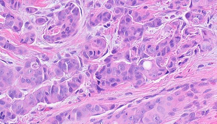
Deliver diagnostic confidence with expertly engineered Leica optics, producing consistently high-quality digital slides that reveal critical details, even in complex cases. Seamlessly integrated into clinical workflows, the Aperio GT 180 DX scanner enables rapid case turnaround, streamlined collaboration, and reproducible results that support timely, informed diagnoses.

Image
Aperio GT 180 Slide Scanner - Proven Technology for Researchers

For Lab Managers

Easy to use and intuitive to learn, the continuous loading, high-automation, and small footprint enables labs to enjoy a smooth, stress-free digital pathology workflow. Advanced feature options such as Manual Scanning*, for challenging slides, and Aperio iQC DX software*, to streamline quality control review, ensure hassle-free, high-quality scans.

Image
Aperio-GT-180-DX-scanner-FOR-LAB-MANAGERS-v2

For IT Managers

Secure, scalable, and easy to integrate; seamlessly connect the Aperio GT 180 DX scanner to your lab’s existing systems and infrastructure and leverage the widely used .svs or standardized DICOM file format for rapid integration with downstream software.

Image
Proven-Technology-for-IT-Managers-700x400

Unrivaled Whole Slide Image Quality

Capture every critical detail with exceptional clarity, from cellular grooves and chromatin structure to intracellular bridges in H&E, or crisp, clear images of your IHC, ISH, and brightfield multiplex slides.

Image
Mitotic Events (Breast)
Image
Intercellular Bridges
Image
Chromatin
Chromatin
Image
Nucleoli
Nucleoli

Precision, Potential to Scale, and Peace of Mind

Digital pathology scan speeds have not always been fast enough at 40x magnification to keep up with high-volume scanning (120k+ slides per year).

Real-Time Focusing (RTF)** offers a potential solution to this problem. It’s a novel method to capture a digital image that combines an imaging line sensor and a focusing line sensor.

  1. While scanning, the focusing line sensor receives focusing data of the tissue.
  2. Proprietary algorithms determine the best focus value.
  3. Then, each best focus value is treated like a control loop with the mechanics, feeding the best focus value to the objective height on the fly.
  4. The positioning adjustments of the objective are performed continuously (in real time) in order to enable the imaging line sensor to capture the image at its best possible focus. Hence, real time focusing.

**US Patent no. 9,841,590

Image
Cellular Groves (Thyroid)

Specifications

Summary of Specifications

Downloads

View all available downloads below

Part/Accessories

PART NUMBER DESCRIPTION

23GT180DXIVD Aperio GT 180 Next Gen Scanner DX IVD
23APERIOSAMSWDX1 Aperio GT DX IVD SAM Software
23GTMANSCANSWDXIVD Aperio GT DX IVD Manual Scan SW Option
23GTEXTFOCSWDXIVD Aperio GT DX IVD Extend Focus SW Option
23GTZSTACKSWDXIVD Aperio GT DX IVD Zstack SW Option

Image
Aperio-GT-180-DX-scanner-640x410