Standard U Frame Compatible
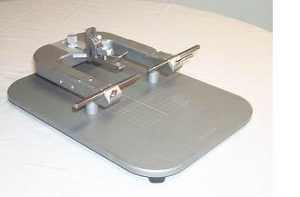
This base without a stereotaxic manipulator holds the anesthetized animal securely in place for procedures not requiring the precision of stereotaxic placement.
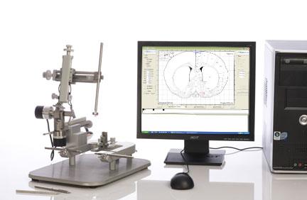
This basic vernier stereotaxic instrumentation offers several practical advantages over competing instruments, and shares these advantages with our digital stereotaxic instruments. The surface finish is both more durable and more easily cleanable than conventional paint
The ear bar locking levers require only one finger to operate, push toward the center. This much eases the task of centering the earbars after installing the animal.
Testimonials
"Bronson Methodist Hospital: a systematic approach to validating xylene‑free processing"

Leica PELORIS improving workflow, safety, and productivity

Excellent results with Leica PELORIS xylene-free processing

Going green with a next generation tissue processor

FAQ
Frequently Asked Questions
To implant bilateral cannulas, one needs only a single manipulator, since this task is done sequentially anyway. To stimulate on the contralateral side, while recording on the ipsilateral side, requires two probes under independent and simultaneous stereotaxic control. This task requires a dual stereotaxic instrument.
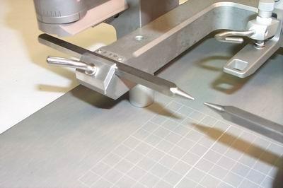
A slightly more stable hold is achieved by the 18° ear bars. However, these puncture the tympanic membrane when installed. The 45° ear bars do not penetrate as far, and do not puncture the tympanic membrane. If hearing acuity could affect the outcome of any planned testing, it is better to use the 45° ear bars. This could include any behavioral or functional testing.
Put one ear bar in blunt end to the center (backwards). Move your probe up close to the flat end, so you can just barely see a gap where light gets through between the earbar and the probe. Operate the vertical drive to raise and lower the probe. The light gap should not change in size. If it does, your probe is not straight in the ML plane. Move the probe around to the side of the ear bar, and repeat the vertical movement to test for straightness in the AP plane. To test if the ML and AP axes are aligned at 90°, move the probe in the ML axes along the ear bar. The gap should stay the same size. If it does not, loosen the locking screw from below the universal joint, and swivel the manipulator until the gap does not appear to change in size as the probe is moved ML.
Next to the ear sockets, there are skull recessions that can feel like the ear holes, but are not as stable. On the rat, grasp the end of the snout and attempt to rock it gently from side to side. If it moves at all, the ear bars are not correctly installed. Swivel the snout up, and let go, or down and let go. If it stays where you leave it and doesn’t move up or down, the ear bars are installed correctly.