
BOND RXm

Helping academic researchers achieve more: BOND RXm – Fully Automated Advanced Staining

Enjoy more time for academic research with BOND RXm fully automating your IHCISH and emerging tests. The unmatched protocol flexibility and fully open reagents mean you retain complete control of your staining. The three independent slide trays also ensure slides run to your timetable.

Contact us to get a quote. Call +1 844 534 2262 to speak with a representative.

The BOND RXm is dedicated to research use only and is not for diagnostic use.

Product Features

Temperature Toolbox

The Temperature Toolbox provides users with the ability to directly edit the temperature of any step (reagent or wash step) in ISH and IHC staining protocols, single or sequential double staining protocols and parallel double staining protocols.

Image
Temperature Toolbox

Time

Now you have extra time to publish

  • Eliminate manual or semi-automated staining
  • Enjoy more time analyzing data, spend less time staining slides
Image
Time

Flexibility

Do it your way, in your space

  • Unmatched protocol flexibility and choice of reagents
  • Fully automated IHCISH and emerging tests
  • A small footprint for benchtop mounting
Image
Flexibility

Consistency

Have more confidence in your results

  • Reduce process variability with full automation
  • Protect tissue with the unique Covertile system
Image
Consistency

BOND RX Software 7.0

BOND RX 7.0 software supports enhanced chromogenic and fluorescent multiplexing functionality by allowing up to 6 individual markers to be visualized on a single slide.

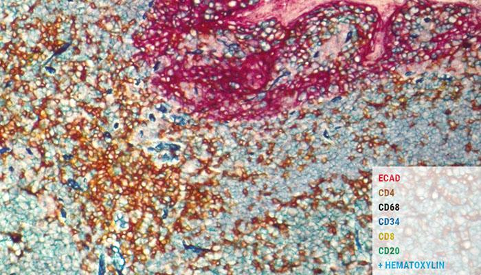
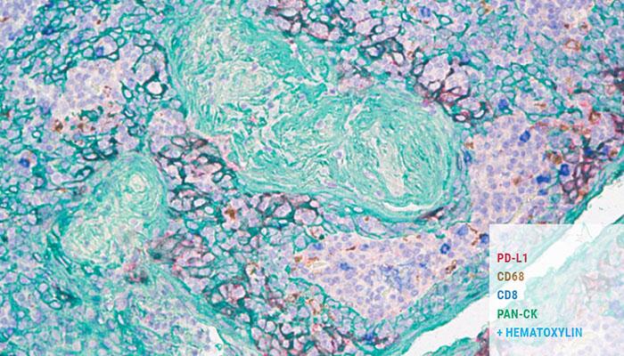
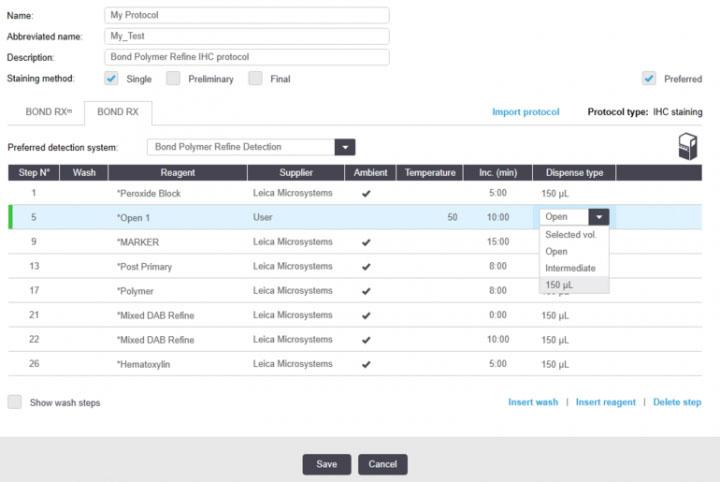
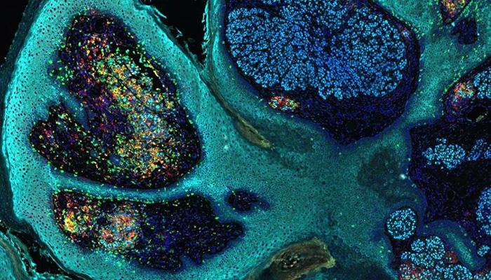
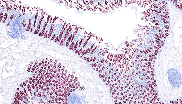
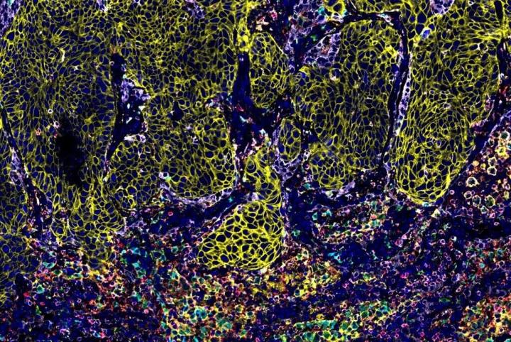
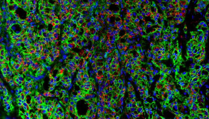
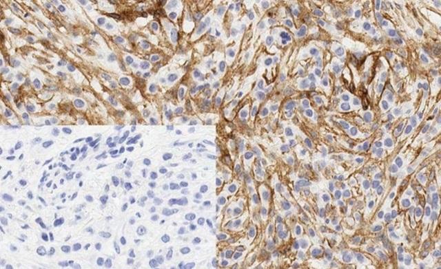
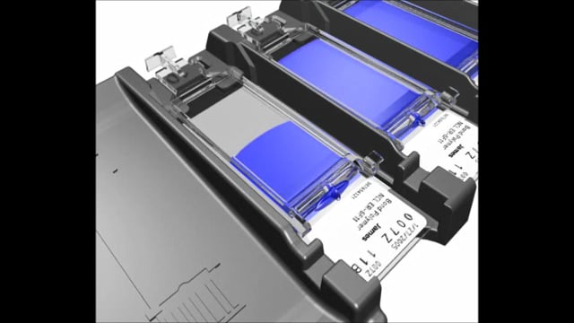
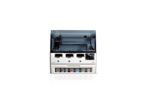
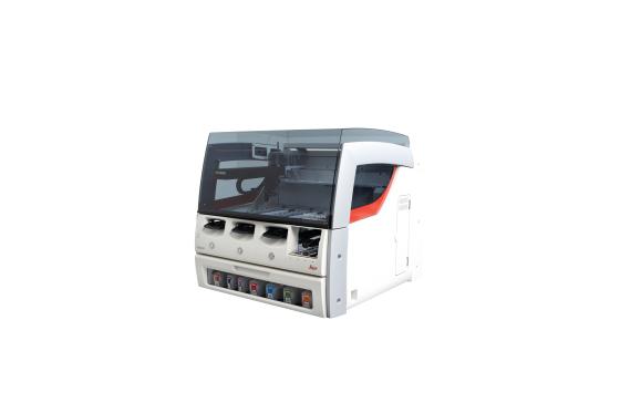
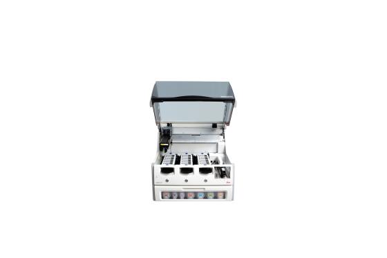
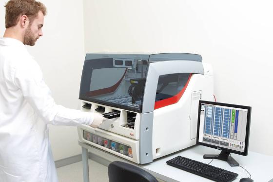
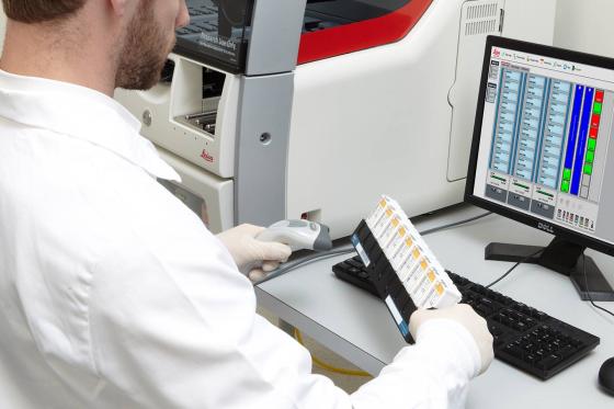
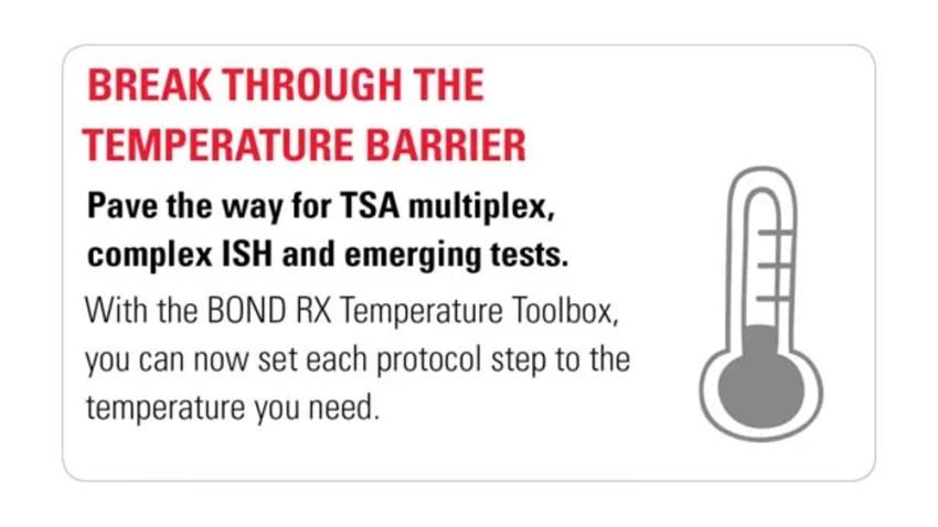
Break Through the Staining Barrier

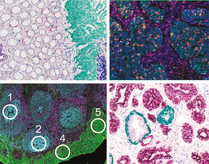
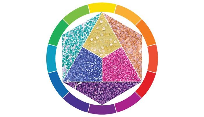
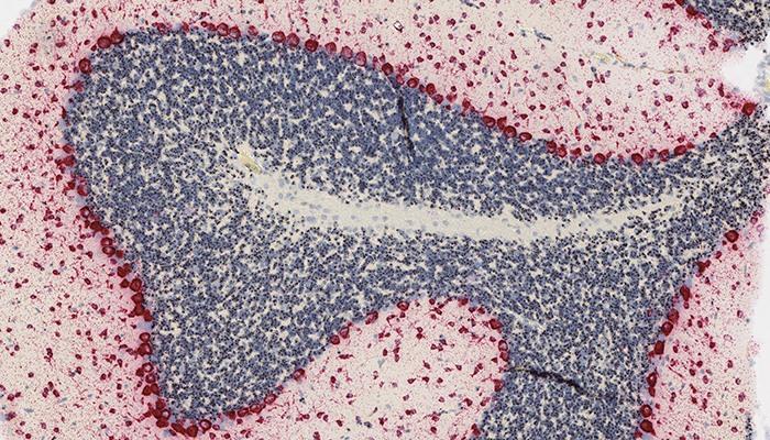
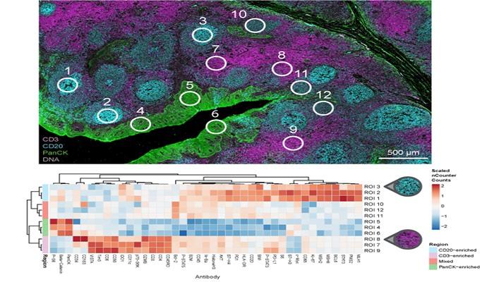
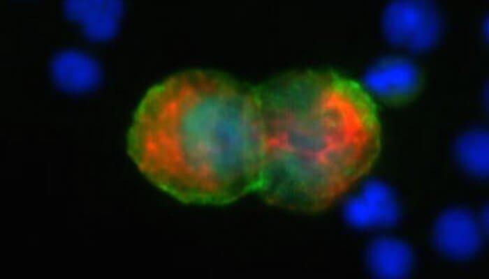
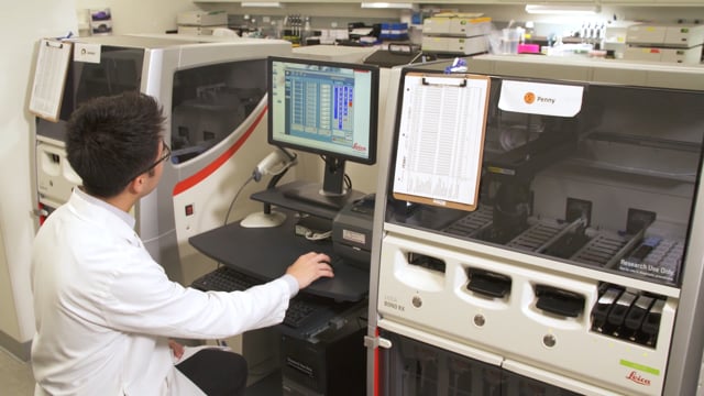
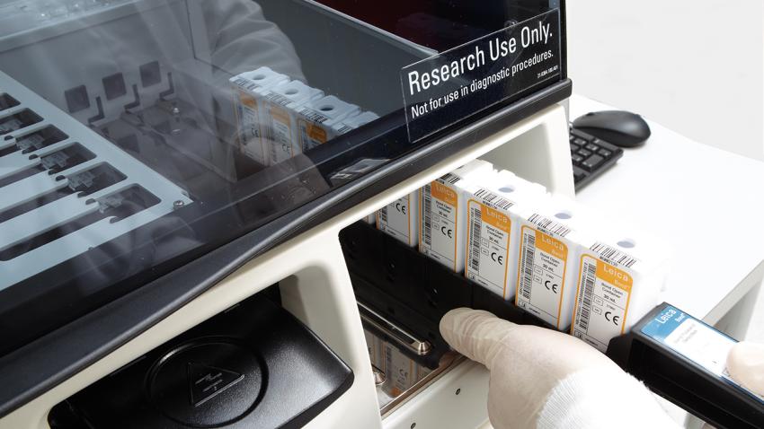
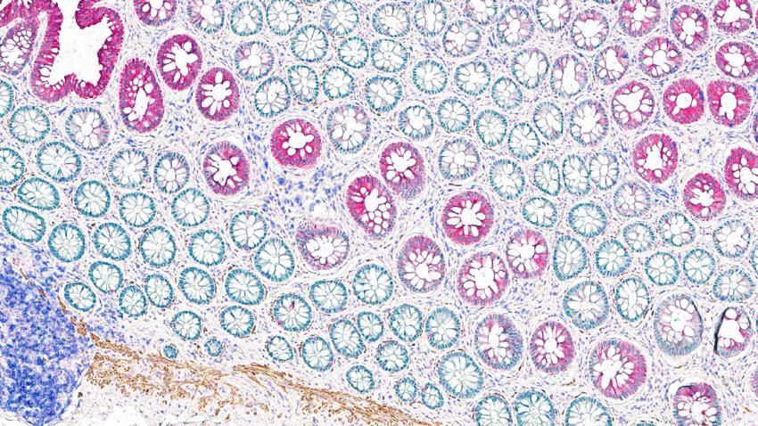
The more markers per slide, the more opportunities to get to your discovery endpoint faster, all while using less precious tissue. Maximize the data you can achieve per sample via fully customized protocols from deparaffinization to counterstain and all the steps in between.

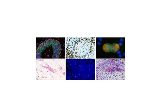
  • Unlock cellular secrets through multiplexing to visualize multiple targets on a single slide
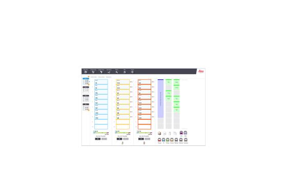
  • Discover the next breakthrough assay by designing your custom detection system and ideal protocol
Image
Break_Through_the_Staining_Barrier

Unlock Cellular Secrets

Multiplex on your research BOND stainer to

  • Discover spatial relationships
  • Determine cellular phenotypes
  • Maximize the data from a single tissue sample

Supported by a wide range of technology partnerships

Image
Unlock_Cellular_Secrets

Design the Next Breakthrough Assay

Freedom to Discover

  • Stain up to 6 markers
  • DNA to RNA to protein
  • FFPE and CTCs
  • Chromogenic, Fluorescent and Sequencing
Image
Design the Next Breakthrough Assay

Explore the Color Options

Choose up to 6 chromogens per slide; plug-and-play or choose your own.

Let the instrument do the mixing with chromogens specifically designed for BOND stainers. With our plug-and-play chromogens, just scan and load on the BOND instrument.

Image
Explore the Color Options

Tailor to Your Target

BOND RX Software 7.0 provides the flexibility to design your ideal protocol

  • Select up to 6 markers per slide
  • Create your own detection systems
  • Fully customize all your research protocols
  • Change time, temperature and dispense type
Image
Tailor to Your Target

Access the Latest Research Advances

Leica Biosystems partners with a range of Open Innovation (OI) partners. As a result, you have many flexible solutions for your research. All protocols and protocol templates developed in conjunction with our OI partners are available on BOND RX and BOND RXm so you can access the latest research advances in the shortest time.

A Fully Integrated RNA ISH Solution
Partner - ACD

Complete up to 30 slides in a single 11-hour run for single plex (and a 14-hour run for duplex) with high-throughput staining. This solution is:

  • Highly specific – Amplifies signals using double z probe strategy and simultaneous background suppression strategy to ensure target-specific binding.
  • Sensitive & Quantitative – Detects single RNA molecules at single cell resolution.
  • Morphological – Provides cell specific expression in information intact tissue architecture.
  • Universal – Use for virtually any gene, species or tissue.

Watch the Partnership Video - ACD RNAscope

Image
A_Fully_Integrated_RNA_ISH_Solution_Partner_ACD

Multiplex Staining With No Intervention
Partner – Akoya*

Free up researchers’ time by creating beautiful, seven-color immunofluorescence slides without any intervention. Up to 30 slides can be created in about 14 hours.

  • Visualize up to six tissue biomarkers at the same time plus a nuclear counterstain.
  • Use the best primary antibodies – regardless of species – with no crosstalk.
  • Optimize the editable protocol with ease so it meets your specific needs.

*Previously PerkinElmer

Image
Multiplex Staining With No Intervention Partner – Akoya

Brilliant, Rapid, Chromogenic Multiplex IHC
Partner - Cell IDx

UltraPlex multiplex chromogenic staining technology is simple, fast and fully automated allowing you to go from staining to analysis in a single day. Generate beautiful images and data on biomarker expression and spatial localization of cell subtypes using the BOND RX System and Aperio Scanners.

  • UltraPlex technology allows the automated detection of multiple markers on BOND RX staining system using primary antibodies from any species, while employing a single antigen retrieval step
    • 3 marker/chromogen stain in 4.5 hours
    • 4 marker/chromogen stain in 6 hours
  • Optimized staining panels of key biomarkers for FFPE tissue
  • Chromogenic detection of cell subtypes coexpressing biomarkers using optimized chromogen combinations
Image
Cell_ID

High Plex, Spatial Profiling of Proteins and RNA from FFPE
Partner – NanoString

Combining standard immunofluorescence techniques with digital optical barcoding technology, the GeoMx™ Digital Spatial Profiler enables high plex, spatially-resolved profiling of protein and RNA. The GeoMx Assays allow for imaging and profiling from a single FFPE or fresh frozen tissue section.

  • Quantify up to 96 proteins and over 1000 RNA targets with spatial context
  • Focus on rare cells or compartment specific regions of interest in tissue
  • Preserve precious samples with non-destructive processing
Image
High Plex, Spatial Profiling of Proteins and RNA from FFPE Partner – NanoString

CTC Analysis For Experimental Pathology Labs
Partner - Rarecyte

The AccuCyte-Cytefinder system integrates with the BOND RX and provides microscopic visualization of circulating tumor cells. It delivers single cell retrieval of taprget cells for downstream genomic analysis.

  • AccuCyte collects nucleated cells including circulating tumor cells from whole blood.
  • CyteSealer makes collection seamless by creating a barrier between the buffycoat and red blood cells.
  • CyteSpreader ensures the precise amount of nucleated cells is spread onto each slide every time.
Image
CTC_Analysis_For_Experimental_Pathology_Labs_Partner_Rarecyte

Single Step Multiplexing, Fully Automated
Partner - Ultivue

Create up to 30, five color, single step multiplexing slides in under 6 hours hours on BOND RX whilst preserving tissue morphology and integrity. Spend less time perfecting difficult manual techniques and more time focusing on your next research breakthrough.

  • Survey and analyse cell phenotypes with spatial context within the tumor microenvironment though high performance tissue-based biomarker multiplex assays
  • Multiplex on a single slide, using a single antibody cocktail and incubation step, powered by DNAbarcoded antibodies with unique, complementary, fluorescent DNA barcodes
  • Preserve sample integrity for additional analyses without damaging tissue or cross talk
  • Image and analyse with Aperio VERSA and Aperio Cellular IF Algorithm
Image
Single_Step_Multiplexing_Fully_Automated_Partner_Ultivue

Product Specifications

DIMENSIONS & WEIGHT

Dimensions (H x W x D): 760 mm x 703 mm x 775 mm
Weight (Dry): 120 kg (265 lbs)
Slide capacity: 30
Reagent container capacity: Titration, 7 mL and 30 mL
Number of reagent containers: 36
Bulk reagent container capacity: 1 L or 2 L
Hazardous waste container capacity: 2L
External bulk waste container capacity: 9L

Downloads

Certificate

Flyer

Infographic

Resources

BOND RTU Antibody and Protocol Data Files

These data files will enable the use of the latest protocol releases for BOND RX/RXm. Local licensing restrictions may apply to some products; please contact your local Leica Biosystems Sales or Support representative should you have any queries. Please ensure the correct data file is downloaded.

Important Notes

  • Do not apply this update to BOND-III or BOND-MAX Systems.
  • Please download and apply the correct update is for your region.
  • We recommend that all BOND systems be updated to the latest software version - BOND RX research software v7.0
  • BOND RX research software v7.0 requires BXD v25 or above
  • Do not apply this update to Systems running RX/RXm v4.0 software; this file is not applicable to software v4.0 Systems.

BOND RXm Publications