
Molecular Instruments

Image
MI_Partner_Logo_800x466

Fully Automated HCR™ Pro RNA-ISH on the BOND RX and BOND RXm Research Staining Platforms by Leica Biosystems

Molecular Instruments® (MI) develops and synthesizes kits powered by its innovative HCR™ platform for bioimaging applications in academic research, drug development, and clinical pathology and diagnostics. MI offers products for automated and manual chromogenic and fluorescence in situ hybridization (ISH) assays, featuring a protease-free workflow, native compatibility with existing IHC/IF assays, and straightforward image analysis.

Features and Benefits of Molecular Instruments’ Technology

Automated RNA Detection with HCR™ Pro RNA-ISH

  • Generate Results in a Single Workday: Stain up to 30 slides in under 10 hours on the BOND RX research staining platform and 11 hours on the BOND RXm research staining platform using pre-loaded protocols.
  • One Protocol, Any RNA: Detect a variety of RNA targets, including microRNAs, splice junctions, lncRNA, and other challenging target types.
  • Plug-and-Play with IHC and IF: Combine HCR™ Pro RNA-ISH with existing IHC and IF protocols on the BOND RX and BOND RXm research staining instruments for simultaneous detection of RNA and protein targets on the same slide.
  • Flexibility with Any Reporter: Order for use with BOND AP- or HRP-driven chromogens.
  • Entirely Protease-Free: Preserve tissue morphology while integrating HCR™ Pro RNA-ISH into existing workflows without additional optimization, enabling easier downstream image analysis and interpretation.

The HCR™ Platform: How it Works

Image
HCR_ Platform Graphics _vertical_660x935

Automated Protein Detection with HCR™ Gold IF

  • Multiplex Same-Species Primary Antibodies with Confidence: Analyze multiple targets from antibodies raised in the same host species without stripping, sequential restaining, or complex workaround workflows.
  • Universal Compatibility with Unmodified Primaries: No antibody conjugation or custom labeling required. You can use standard, off-the-shelf primaries and keep your existing antibody portfolio and validation work intact.
  • Reduce Hands-On Complexity: Add a short room-temperature encoding step, then run automated staining on the BOND RX research staining platform with minimal added workflow burden.
  • Build Richer Spatial Protein Profiling on a Single Slide: Preserve tissue context while simplifying multiplex assay design, with simultaneous detection of up to five protein targets.

Research Focus

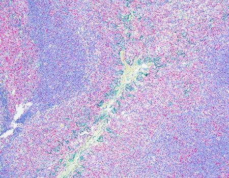
From RNA to Protein: Understanding PD-L1 Expression in Tonsillar Immune Microenvironments with HCR™ Pro RNA-CISH and IHC

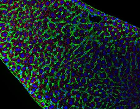
HCR™ Pro RNA-ISH enables high-resolution visualization of RNA expression within tissue sections, offering a powerful tool for studying immune cell dynamics. Here, PD-L1, a key immune checkpoint ligand, is explored as both an RNA (imaged in red) and protein (imaged in green) marker to investigate its expression patterns in normal human tonsil tissue.

 With its diverse immune cells and organized lymphoid structure, the normal tonsil is an ideal model for studying immune regulation. Detecting PD-L1 at both the RNA and protein levels within the same sample reveals the interplay between transcriptional activity and protein expression, shedding light on this critical marker’s role in immune homeostasis. 

This integrated RNA and IHC approach maximizes the use of valuable samples, enabling a comprehensive analysis of PD-L1’s spatial expression and functional dynamics. Such insights are essential for advancing our understanding of immune checkpoint biology in disease, paving the way for the development of more effective, targeted immunotherapies.

Image
HCR_Pro_RNA_CISH_IHC_PD_L1_Partner_Page_Research_Application_1_450x350

Protease-Free HCR™ Pro RNA-ISH: Preserving Tissue Architecture for Seamless IHC/IF Co-detection

One of the key advantages of HCR™ Pro RNA-ISH is its protease-free nature, which preserves tissue morphology while maintaining high specificity for RNA detection. This makes it ideally suited for seamless integration with IHC and IF assays, allowing researchers to co-detect RNA and protein markers within the same tissue section, without compromising structural integrity. 

In this example, the use of an HCR™ Membrane Stain (imaged in green) highlights the precise segmentation of cellular membranes, enabling clear and distinct visualization of Ppib RNA (imaged in red) expression patterns along the cell boundaries. By combining HCR™ Pro RNA-ISH with IHC/IF assays, researchers can gain a deeper understanding of cellular interactions and gene expression patterns.

Image
HCR_Pro_RNA_FISH_IF_Membrane_Stain_Partner_Page_Research_Application_2_450x350

Automated Multiplex IF and Spatial Analysis for Colorectal Tumor Microenvironment Profiling

This AACR collaboration with Leica Biosystems, Molecular Instruments, Abcam, and Indica Labs highlights an end-to-end workflow for profiling the colorectal tumor microenvironment in FFPE tissue. On the BOND RX research staining system, Abcam antibody clones were first evaluated in single-plex format using HCR™ Pro IHC and shown to produce staining patterns comparable to BOND Polymer Refine Detection on serial sections. Building on that validation, HCR™ Gold IF enabled same-species multiplex immunofluorescence without iterative staining, supporting flexible panel development while preserving tissue morphology.

Using HALO® for tissue segmentation, cell phenotyping, density measurements, and spatial analysis, the workflow distinguished healthy colon from colorectal cancer based on epithelial organization, immune cell distribution, and proliferative activity. Multiplex analysis of panCK, Ki67, CD3, and CD68 showed enrichment of tumor and proliferating tumor cells in CRC epithelium, with immune populations more abundant in stromal regions and altered T-cell localization at the tumor- stroma interface. Together, the poster shows how automated HCR™ workflows plus quantitative image analysis can support scalable, spatially resolved tumor microenvironment studies.

DOWNLOAD NOW

Image
automated-flexible-multiplex-immunofluorescence-for-Tumor-Microenviroment-Profiling-Using-HCR

Meet the Molecular Instruments Team

Image
Harry_Choi_450x450

Dr. Harry M.T. Choi

Founder & CEO

Harry, co-founder and CEO of MI, led 3 generations of HCR™ technology development at Caltech, exploiting new concepts from dynamic nucleic acid nanotechnology to overcome challenges to multiplexed quantitative bioimaging in thick, autofluorescent samples.

Image
Aneesh_Acharya_450x450

Dr. Aneesh Acharya

Chief Operating Officer

Aneesh is the Chief Commercial Officer at MI, bringing both commercialization experience from his work in the tech industry, but also technical expertise from his graduate work at Caltech developing platform technologies now used across HCR™ Gold and Pro products.

Brochure on HCR ™ Assays on the BOND RX Research Staining Instrument
 

Image
1218-GBL-EN-v1-MI-x-LBS-Brochure-cover

Download Now

For Research Use Only. Not for Use in Diagnostic Procedures