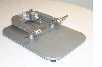
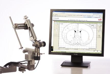
Angle Two Small Animal Stereotaxic Instrument Stereotaxic Instrument with Atlas Integration in Computer
The Angle Two small animal stereotaxic instrument enables varying the angle of approach between surgeries without loss of accuracy. Target setting and path of approach are integrated with an onscreen atlas, either mouse or rat or both. Virtual Skull Flat feature corrects for head tilt. Spend less time on each animal, with more accurate results, and vary the angle of approach to avoid confounding action-at-target with path-of-approach.
With no verniers to read or arithmetic to do, risk of human error is greatly reduced. The neuroscientist can improve the rigor and interpretability of stereotaxic research without adding heavy calculations or risk of error to the protocol, and therefore design experiments with fewer animals.
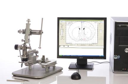
Includes configured computer with software for your choice of mouse or rat installed, manual fine drive for precise transit and positioning. Installed encoders connect to computer allow it to keep track of electrode tip location once shown the position of Bregma.
Learn more
Fewer animals needed
Reduced risk of human error, and accuracy enhancing features in the software, result in fewer misses so no wasted animals and resulting wasted testing.
Vary angle of approach with no loss of accuracy
Every surgery can be done from a different angle, with no more time required and no loss of accuracy. Avoid confounding of path-of-approach with action-at-target.
Includes on-screen atlas
Click on atlas to define target coordinates. Visual display of path to be followed through brain.
Virtual skull-flat
Instrument measures head tilt from skull flat, and mathematically corrects target position coordinates. No need for trial and error head tilt adjustments.
Save and recall coordinates sets
For multiple user labs, store the coordinate set in an on-screen icon at the bottom. Recall by clicking on the icon when in Set Target mode.
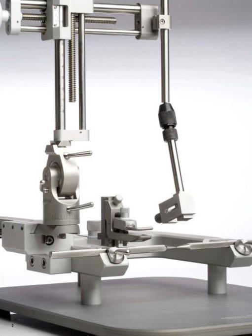
Manual Fine Drive
Low mounted manual fine drive on the DV axis helps avoid hand transmitted vibration during operation of the DV drive, and makes it easy to avoid overshoot.
Testimonials
"Bronson Methodist Hospital: a systematic approach to validating xylene‑free processing"

Leica PELORIS improving workflow, safety, and productivity

Excellent results with Leica PELORIS xylene-free processing

Going green with a next generation tissue processor

Downloads
IFU and SDS
Publications
The following publications cite the Leica Biosystem’s Angle Two Stereotaxic instrument for Research Use Only.
- Sanathara, N. M., Garau, C., Alachkar, A., Wang, L., Wang, Z., Nishimori, K., … Civelli, O. (2018). Melanin concentrating hormone modulates oxytocin-mediated marble burying. Neuropharmacology, 128, 22–32. doi.org/10.1016/J.NEUROPHARM.2017.09.008
- Liu, G., Swanson, J., Pekarek, B., Panneerselvam, S., Ung, K., Tepe, B., … Arenkiel, B. R. (2018). A Combinatorial Approach to Circuit Mapping in the Mouse Olfactory Bulb. doi.org/10.1007/978-1-4939-7549-5_7
- Ghazale, H., Ramadan, N., Mantash, S., Zibara, K., El-Sitt, S., Darwish, H., … Soueid, J. (2018). Docosahexaenoic acid (DHA) enhances the therapeutic potential of neonatal neural stem cell transplantation post—Traumatic brain injury. Behavioural Brain Research, 340, 1–13. doi.org/10.1016/J.BBR.2017.11.007
- Gelegen, C., Miracca, G., Ran, M. Z., Harding, E. C., Ye, Z., Yu, X., … Franks, N. P. (2018). Excitatory Pathways from the Lateral Habenula Enable Propofol-Induced Sedation. Current Biology, 28(4), 580-587.e5. doi.org/10.1016/J.CUB.2017.12.050
- López, A. J., Jia, Y., White, A. O., Kwapis, J. L., Espinoza, M., Hwang, P., … Wood, M. A. (2018). Medial habenula cholinergic signaling regulates cocaine-associated relapse-like behavior. Addiction Biology. doi.org/10.1111/adb.12605
- Schroeder, M., Drori, Y., Ben-Efraim, Y., Metabolism, A. C.-M., & 2018, U. (2018). Hypothalamic miR-219 regulates individual metabolic differences in response to diet-induced weight cycling. Elsevier, 9, 176–186. Retrieved from www.sciencedirect.com/science/article/pii/S2212877817306725
- Hagit, C., Ella, V., Kaplan, Z., Joseph, Z., & Aleksander, M. A. (2017). Neuropeptide S in the basolateral amygdala mediates an adaptive behavioral stress response in a rat model of posttraumatic stress disorder by increasing the expression of BDNF and the neuropeptide YY1 receptor. European Neuropsychopharmacology. doi.org/10.1016/J.EURONEURO.2017.11.006
- Ramot, A., Jiang, Z., Tian, J.-B., Nahum, T., Kuperman, Y., Justice, N., & Chen, A. (2017). Hypothalamic CRFR1 is essential for HPA axis regulation following chronic stress. Nature Neuroscience, 20(3), 385–388. doi.org/10.1038/nn.4491
- Schiff, H. C., Bouhuis, A. L., Yu, K., Penzo, M. A., Li, H., He, M., & Li, B. (2017). An insula-central amygdala circuit for behavioral inhibition. BioRxiv, 156216. doi.org/10.1101/156216
- Seo, J.-S., Wei, J., Qin, L., Kim, Y., Yan, Z., & Greengard, P. (2017). Cellular and molecular basis for stress-induced depression. Molecular Psychiatry, 22(10), 1440–1447. doi.org/10.1038/mp.2016.118
- Namestnikova, D., Gubskiy, I., Kholodenko, I., Melnikov, P., Sukhinich, K., Gabashvili, A., … Yarygin, K. (2017). Methodological aspects of MRI of transplanted superparamagnetic iron oxide-labeled mesenchymal stem cells in live rat brain. PLOS ONE, 12(10), e0186717. doi.org/10.1371/journal.pone.0186717