
From Microscope To Monitor A Proprietary Color Match Method from Leica Biosystems


Digital pathology is quickly growing across the globe as it offers increasing benefits to standardize and optimize the pathology lab workflow. Pathologists can now retrieve their cases in digital form and review and sign off cases using a PC and monitor. However, digital pathology companies offer many different monitors, depending on the company. Pathologists are often frustrated by the color variation of a digital image as it may not match the microscope color experience They often prefer a color that looks as close as possible to the typical experience using a microscope.
How can we color match the image viewed on the monitor to what is seen in the microscope?
The problem is that no company had calibrated the color of the histology stain to the color of the monitor and color of the whole slide image. Our innovation team worked together to solve this issue.
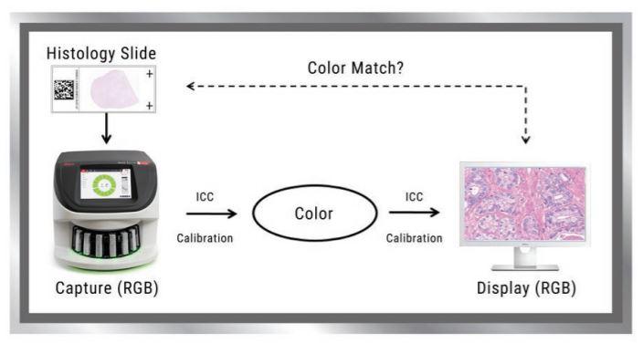
Image Capture RGB values versus Display (Monitor) RGB values
When an Aperio digital pathology scanner scans a slide it produces a whole slide image that contains calibrated red-green-blue (RGB) values because the image is a color image. When the image is viewed on a monitor, the monitor also has its own separate calibrated RGB values (for example sRGB calibration by the manufacturer) because it is a color monitor. The problem is that image capture RGB values generated during image acquisition do not match the monitor display RGB values.
Many customers simply assume image capture RGB values will match the monitor RGB values. But this is not the case.
A novel solution to calibrate color using Histology slides (patent pending).
The goal is to reproduce what is seen in the microscope so that the same information is presented to the pathologist. In order to do this, the scanner and monitor need to be matched properly in order to produce accurate color. Leica Biosystems innovation solved this by creating a novel method, which includes the following:
- Input color is measured on a histology slide using a patented spectral method (standard D50 light used in microscopes).
- Output color is measured on the monitor display. Small patches of a whole slide image are magnified on the monitor and a colorimeter/spectrophotometer is used to measure the color of light for each patch.
- The scanner and monitor display are then calibrated separately for accurate color.
- An ICC profile is created to convert between RGB values across scanner and monitor to produce accurate color.
- Each monitor brand and model has its own ICC profile, and Aperio viewers calibrate image color against that ICC profile for consistent visualization.
Pathologists Benefit as Monitor Color Matches Histology Slides and Microscope
As digital pathology ramps up its adoption across the globe, it is important to consider the color of the image that is displayed on the monitor as Pathologists prefer that the color match the microscope (D50 light passing histology stained slides). By utilizing Leica Biosystems novel calibration methodology an ICC profile has been created to convert between RGB and color so that when a Pathologist views the image on a monitor it matches the color to histology stained slides.
For Research Use Only. Not for Use in Diagnostic Procedures
About the presenters
Dr. Allen Olson is a Technical Lead Engineer and Scientist at Leica Biosystems and has been with the company since the early days of Aperio. His subject matter specialties include algorithms for automating whole slide scanning and color calibration of scanner and display. He holds a PhD in Earth Sciences (Geophysics) from Scripps Institution of Oceanography and a BA in Applied Mechanics and Engineering Sciences from the University of California San Diego. Dr. Olson is co-inventor on numerous patents related to whole slide imaging and color.
Lance Mikus earned his bachelor’s degree from UCLA. He worked as a scientist and lab manager at the UW-Madison Medical School, resulting in several publications, and went on to earn an MBA focused on Entrepreneurship and Marketing from the UW-Madison School of Business. Mr. Mikus has 20 years of commercial operations experience in the healthcare industry. He also holds a green belt in six sigma quality practices, is a certified practitioner in launch excellence and has been awarded 5 patents.
Related Content


Leica Biosystems content is subject to the Leica Biosystems website terms of use, available at: Legal Notice. The content, including webinars, training presentations and related materials is intended to provide general information regarding particular subjects of interest to health care professionals and is not intended to be, and should not be construed as, medical, regulatory or legal advice. The views and opinions expressed in any third-party content reflect the personal views and opinions of the speaker(s)/author(s) and do not necessarily represent or reflect the views or opinions of Leica Biosystems, its employees or agents. Any links contained in the content which provides access to third party resources or content is provided for convenience only.
For the use of any product, the applicable product documentation, including information guides, inserts and operation manuals should be consulted.
Copyright © 2026 Leica Biosystems division of Leica Microsystems, Inc. and its Leica Biosystems affiliates. All rights reserved. LEICA and the Leica Logo are registered trademarks of Leica Microsystems IR GmbH.