Proven Technology for Researchers
Effortless scanning, exceptional results with excellent image quality, innovative technology and seamless integration – all driven towards faster turnaround time and accuracy for your important projects
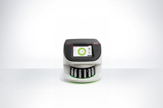
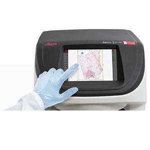
The Aperio GT 450 scanner combines advanced technology and innovation, providing flexible scanning and automated workflows for efficient, high-quality results.
For Research Use Only. Not for use in diagnostic procedures.
*Scan speed assumes 15mm x 15mm area at 40x
** Optional features available to meet your workflow requirements. Some features not available in all countries.
Aperio is a trademark of the Leica Biosystems group of companies in the USA and optionally in other countries. GT and GT 450 are trademarks of Leica Biosystems Imaging, Inc. Other logos, product and/or company names might be trademarks of their respective owners.

Effortless scanning, exceptional results with excellent image quality, innovative technology and seamless integration – all driven towards faster turnaround time and accuracy for your important projects


Delivering researchers with an efficient solution for variability in slide preparation, Manual Scan provides:
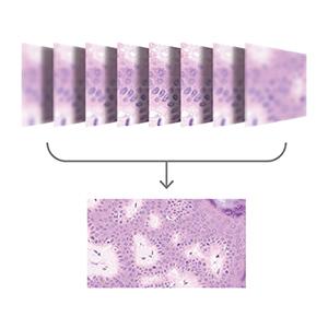
Experience an efficient imaging solution that improves image quality while reducing storage needs. Extended Focus features include:
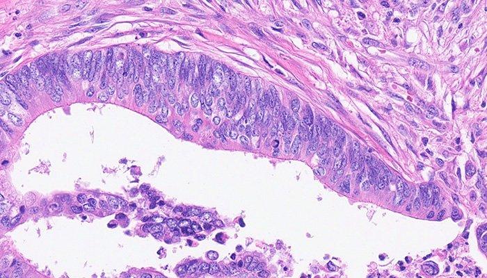
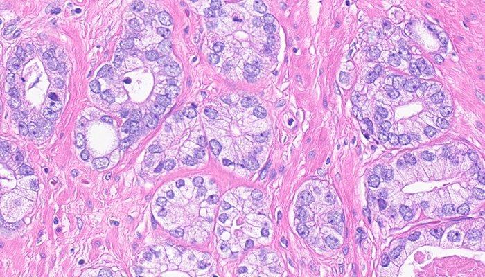
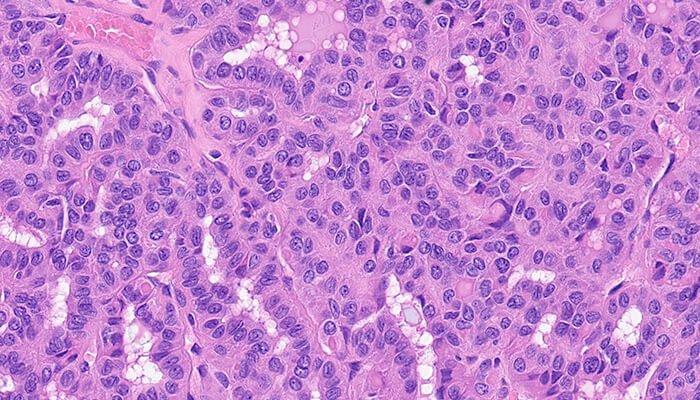
Aperio GT 450 uses a high-performance objective manufactured by Leica Microsystems, which has produced world-class optics since 1847. Leica optics deliver exceptional image quality
Deliver rapid results with confidence.
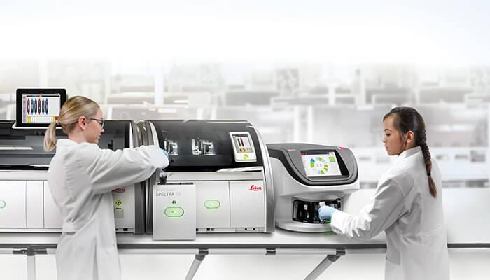
The Aperio GT 450 is simple to operate and helps you complete scanning quickly and easily, so you can focus on other tasks in the lab: 15 racks of 30 slides (450 slides total) can be loaded directly from HistoCore SPECTRA CV Coverslipper into the Aperio GT 450. The slides are then automatically scanned at 81 slides per hour at 40x for a 15mm x 15mm area.
Providing lab technicians with an intuitive and efficient scanning solution, the benefits of Manual Scan include:
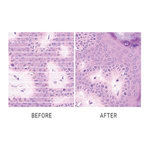
The Aperio GT 450 Default Calibration Point reduces the chance of striping from the start, allowing lab managers to maintain image clarity and avoid corrective measures. This ensures consistently high-quality results with no extra effort.
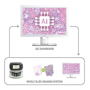
Optional Aperio iQC software enhances image quality control, ensuring confidence before scans are delivered to the researcher.
The Aperio GT 450 offers fast, secure, and flexible IT architecture. A centralized Scanner Admin Manager (SAM) server and dedicated software provides you with a data management solution that can remotely set up and monitor multiple Aperio GT 450s at a time.*
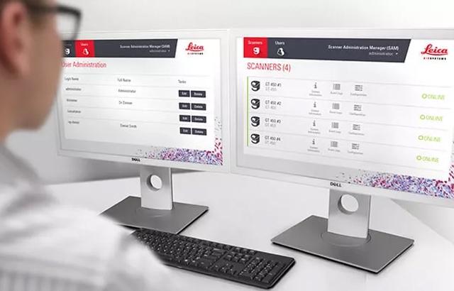
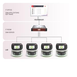
No more workstations needed. A centralized Scanner Admin Manager (SAM) enables IT professionals to scale up Digital Pathology operations securely and efficiently. SAM server and software allow IT managers to set up and monitor Aperio GT 450 scanners via the network, including dashboard status views, PIN controls and log out times.
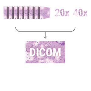
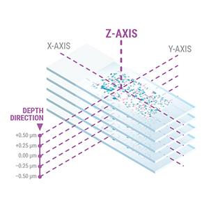
The DICOM format enhances compatibility for Z-Stacking, 20x and 40x magnification, offering high-quality images that can be easily shared and accessed across different systems. Now available alongside .svs, DICOM enables image clarity, improved collaboration and a streamlined workflow.
(for best viewing results we recommend the Aperio Viewing Station which includes a calibrated monitor)
Digital pathology scan speeds have not always been fast enough at 40x magnification to keep up with high-volume scanning (120k+ slides per year). Real-Time Focusing (RTF)** offers a potential solution to this problem. It’s a novel method to capture a digital image that combines an imaging line sensor and a focusing line sensor.
**US Patent no. 9,841,590
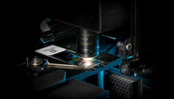
The high-performance objective included with the Aperio GT 450 is specifically designed to maximize field of view for high speed digital pathology scanning. Most objective are designed similar to the human eye: round, which focuses light in the center and limits field of view. The objective on the Aperio GT 450 includes an extra-wide flat field correction that enables a much larger field of view (1mm) that can accommodate extremely large digital images for fast scans and 0.26 um/pixel resolution at 40x magnification*.
*Available in 20x magnification.
From tissue processing to Digital Pathology, our solutions empower labs with efficiency, precision, and reliability.
Aperio GT 450 Scanner - Manual Scan

Introducing the Aperio GT 450