Whole-Slide-Imaging-hero-banner

Whole Slide Imaging

How Whole Slide Imaging is changing pathology

Whole Slide Imaging (WSI) is revolutionizing the field of pathology, ushering in an era of unprecedented access to Image quality, ease of use, and speed. WSI allows for the digital acquisition and storage of tissue slides and has immense practical implications for researchers.

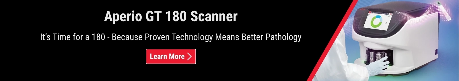
What is Whole Slide Imaging?

Whole Slide Imaging involves digitally scanning a tissue slide containing thin sections of tissue specimens for microscopic examination and storing it as digital images. This process allows for remote collaboration and analysis that can care about Image quality, ease of use, and speed.

How Does Whole Slide Imaging Work?

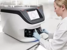
The process begins with the selection of an appropriate scanner and microscope. Once these have been chosen, slides are prepared according to standard protocols. The slides are then placed on the scanner’s stage and scanned using a motorized platform that moves across the surface at varying speeds depending on the resolution required. The image data accumulated during scanning is then processed using unique algorithms to create a digital version of the specimen that can be stored in a database. This image data can be shared with colleagues for collaboration and analysis.

Image
What-is-Whole-Slide-Imaging-850x600

Advantages of Whole Slide Imaging in Pathology

Whole Slide Imaging has numerous potential benefits for research professionals

Image
Advantages-of-Whole-Slide-Imaging-in-Pathology-500x450.jpg

Increased Efficiency in Pathology Workflow
With WSI researchers can review slides remotely without having to physically handle them first which means ease of use. Furthermore, since WSI does not require any special preparation before scanning, there is no need for reagents or other consumables, which could be cost-efficient.


Remote Reviewing & Analysis
Digital versions of slides stored in databases allow researchers from different institutions worldwide to collaborate from anywhere with an internet connection. 


Digital Archiving & Accessibility
WSI enables researchers to obtain objective measurements from images such as cell counts, size distributions, cellular patterns, and staining methods. 
 

Challenges & Limitations of Whole Slide Imaging

Whole Slide Imaging (WSI) may have immense potential and could improve research outcomes. However, integrating WSI with existing research infrastructure, ensuring standardization, and managing and analyzing large volumes of data can pose challenges.

 Integrating WSI with existing research workflows and infrastructure

While WSI has potential for to streamlining workflows, and improving research outcomes, integration with existing infrastructure can pose a challenge. Organizations that have invested in traditional microscopy infrastructure may be reluctant to switch to WSI, as it may require specialized equipment and software. Furthermore, digitizing existing slides may be time-consuming, resource intensive and can pose challenges to maintaining data.

 Standardization and quality control

As with any imaging technology, ensuring consistent image quality and reproducibility can be crucial for research applications. Challenges surrounding standardization and quality control include variations between imaging platforms, differences in staining techniques, and the potential for technical errors in digitizing and processing images. Addressing these challenges will be critical for enabling WSI as a reliable and reproducible research tool. 

Data management and analysis

WSI generates large volumes of data, which can mean significant s storage, organization, and analysis challenges. Handling and analyzing these data can require specialized software and computing infrastructure, which could mean adding further complexity to research workflows. Data security and privacy concerns may also arise when working with WSI data. Effectively managing and analyzing WSI data will be critical for realizing the full potential of this technology for research applications.

Research Organizations 

Whole Slide Imaging has enabled the Contract Research Organization (CRO), Pharma, and Biobanks departments to digitize their microscope slides quickly and accurately, making them easier to store and share with other research professionals. This enhanced access to tissue slides also allows for rapid analysis. Furthermore, due to its ability to enable remote viewing between researchers over long distances, whole slide imaging has been embraced by research organizations. It may be a cost-effective solution that can improve collaboration between research professionals while preserving quality standards.

Contract Research Organization

This technology has allowed analyzing glass slides more quickly while simultaneously creating digital images that can be stored electronically and shared with various stakeholders. Additionally,  using automated image analysis algorithms powered by artificial intelligence (AI) can free up research staff for other tasks.

Another significant benefit of WSI is that it can improve scalability. With digitization comes the ability to rapidly expand operations without needing additional physical resources such as additional buildings or staff members. Furthermore, large data sets can be centralized into one server without taking up too much physical space or requiring extra personnel, allowing research staff more efficient access to information. Additionally, WSI  provides more widespread access to data from various sources due to features, such as teleconferencing, making it easier to connect remotely with colleagues across all geographical boundaries.

Image
Contract-Research-Organization-640x410

Pharmaceutical and Biotech Companies

Whole Slide Imaging (WSI) can improve research and development process for pharmaceutical and biotech companies. WSI could allow for high-resolution imaging of tissue specimens, enabling researchers to observe and analyze molecular changes within the tissue at a microscopic level. By digitizing tissue samples, WSI could allow remote access to high-quality images, enabling researchers to work collaboratively on projects even when they are not physically present in the lab. The rise of digital pathology and WSI may bring profound changes in the life sciences field.

Overall, Whole Slide Imaging represents a great breakthrough in research technology and can hold immense potential for pharmaceutical and biotech companies looking to improve the way they conduct research and development.

Image
Pharmaceutical-and-Biotech-Companies-640x410-3

Tissue Biobanking and Research Institutions

Whole Slide Imaging allows researchers at tissue biobanking institutions access to digital image archives containing hundreds or thousands of individual slides. These digital images can be used as part of ongoing studies into diseases or treatments without needing additional physical specimens or costly lab equipment.

The slides are easier to distribute among different research groups than traditional glass slides, plus they offer better protection against environmental factors like dust or humidity that could interfere with their quality over time. Additionally, they provide more precise information regarding subcellular changes, which aids the development of new drugs or therapies that may be more effective than existing ones due to their specificity at a molecular level.

Image
Tissue-Biobanking-and-Research-Institutions-640x410-1

Future Developments and Trends in Whole Slide Imaging

Whole Slide Imaging technology continues to embraced the field of pathology looking for efficient and remote collaboration between researchers. As this technology evolves, it is expected to make significant contributions to the life sciences and research fields. In the future, advancements in Whole Slide Imaging could focus on enhancing the resolution and accuracy of digital images. Developers are exploring the use of artificial intelligence and machine learning to assist in the analysis of digital images.  The development of Whole Slide Imaging technology can contribute to advancements in life science research.

For Research Use Only. Not for use in diagnostic procedures.


Related content:

David S. McClintock, Jacob T. Abel & Toby C. Cornish. “Whole Slide Imaging Hardware, Software, and Infrastructure” (2021) available here

Ole-Johan Skrede, Sepp De Raedt, Andreas Kleppe, Tarjei S Hveem, Knut Liestøl, John Maddison, Hanne A Askautrud, Manohar Pradhan, John Arne Nesheim, Fritz Albregtsen, Inger Nina Farstad, Enric Domingo, David N Church, Arild Nesbakken, Neil A Shepherd, Ian Tomlinson, Rachel Kerr, Marco Novelli, David J Kerr, Håvard E Danielsen, “Deep learning for prediction of colorectal cancer outcome: a discovery and validation study” (2020) Available here

Deep learning for prediction of colorectal cancer outcome: a discovery and validation study,
TECHNOLOGY AND CODE article, “Deep Learning-Based Mapping of Tumor Infiltrating Lymphocytes in Whole Slide Images of 23 Types of Cancer” (2022) Available here

Liron Pantanowitz, Paul N. Valenstein, Andrew J. Evans, Keith J. Kaplan, John D. Pfeifer, David C. Wilbur, Laura C. Collins, Terence J. Colgan, “Review of the current state of whole slide imaging in pathology” Journal of Pathology Informatics, Volume 2, Issue 1, 2011, 36, ISSN 2153-3539. Available here

Leica Biosystems - Whole Slide Imaging Solutions

Digital Pathology

Adopting digital pathology is complex, but it doesn’t have to be complicated. Leica Biosystems can clarify your organization’s path to digitally driven discoveries or diagnostics. Our world-leading, modular technology is supported by experience with thousands of Aperio Digital Pathology implementations worldwide.

Digital Pathology Solutions