
Educational Portal
The Life Science Portal is a repository of educational content that is especially curated for the research community. Discover resources on Histology techniques and Pre-Analytics, Multiplex and ISH Applications, and Digital Pathology Imaging and Scanning.

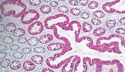
Histology & Pre-Analytics
Ensuring pre-analytic quality in histology drives high-caliber data in downstream applications. Explore our webinars, articles, and resources for improving your processes.
IHC & Multiplexing
Researches need clear results to discover new treatments. BOND RX fully automated research stainers provide the flexibility your need to explore new possibilities, accurate results to ensure nothing is missed, and rapid, cost-effective operation, so you can perform more tests.

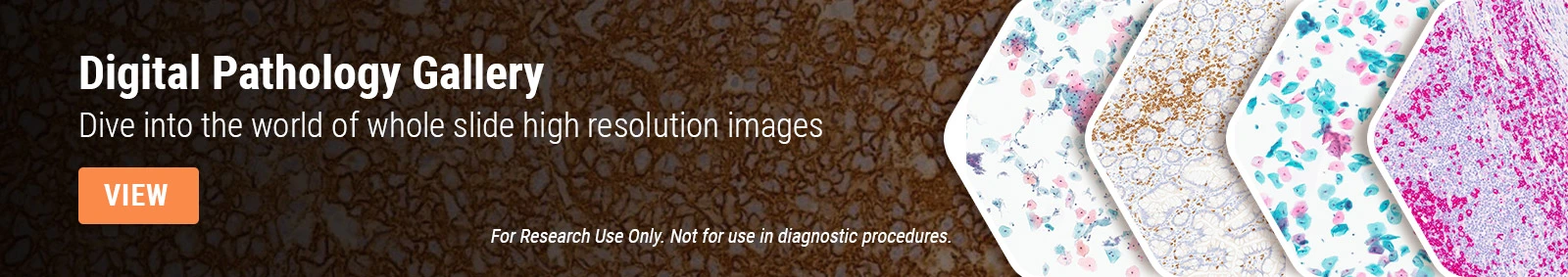
Digital Pathology Imaging & Scanning
Our digital slide scanning products offer unprecedented image quality, speed and reliability for whole slide imaging; making Aperio Digital Pathology scanners the optimal choice for research professionals.
Resource Portal
The Life Science Portal is an evidence-based platform for researchers to gain insights into cutting-edge technologies and investigate how practitioners are implementing them. Read about Innovation Partners and Technologies, Stain Galleries, Peer-Reviewed Publication Repositories, and Life Science Applications.
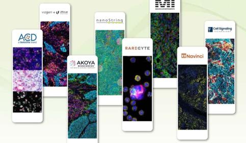
Innovation Partners & Technologies
Technologies partnered with a fully automated workflow yield visionary outcomes.
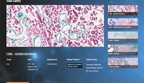
Stain Gallery
Explore the virtual stain gallery with images showing different staining techniques, such as multiplex biomarker detection in tissue samples performed on the BOND RX fully automated stainer.

Peer-Reviewed Publications Repository
The Leica Biosystems Life Science peer-reviewed publication repository offers a method for building a bibliography of scientific publications referencing our products.

Life Sciences Applications
Accelerate Your Journey, Imagine The Possibilities
Leica Biosystems research portfolio provides instruments, solutions, and support for each step of your tissue-based journey; for biomarker to digital analysis, and beyond.
Featured Life Sciences Products
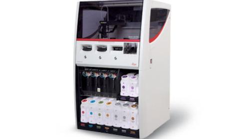
BOND RX
The BOND RX high-throughput research stainer delivers speed and flexibility for IHC, ISH, and multiplexing staining with either chromogenic or fluorescence markers.
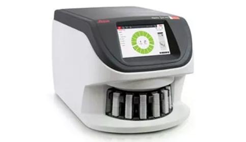
Aperio GT 450 Scanner
Proven technology, with enhanced features, provides efficient workflows and excellent image quality, ensuring seamless integration and secure, optimized delivery of your research.
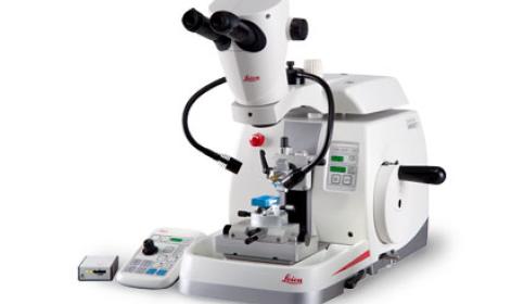
HistoCore NANOCUT R
When your microtomy involves diverse samples in order to discover new breakthroughs in your research projects, then NANOCUT R is the optimal solution for you.
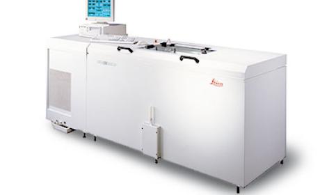
Leica CM3600 XP
Fully computerized cryomacrotome for whole‐body sectioning of small animals.
Subscribe to our mailing list to learn about our upcoming symposiums and new research-focused product launches.
FOR RESEARCH USE ONLY. Not For Use In Diagnostic Procedures.
Leica Biosystems content is subject to the Leica Biosystems website terms of use, available at: Legal Notice. The content, including webinars, training presentations and related materials is intended to provide general information regarding particular subjects of interest to health care professionals and is not intended to be, and should not be construed as, medical, regulatory or legal advice. The views and opinions expressed in any third-party content reflect the personal views and opinions of the speaker(s)/author(s) and do not necessarily represent or reflect the views or opinions of Leica Biosystems, its employees or agents. Any links contained in the content which provides access to third party resources or content is provided for convenience only.
For the use of any product, the applicable product documentation, including information guides, inserts and operation manuals should be consulted.
Copyright © 2024 Leica Biosystems division of Leica Microsystems, Inc. and its Leica Biosystems affiliates. All rights reserved. LEICA and the Leica Logo are registered trademarks of Leica Microsystems IR GmbH.