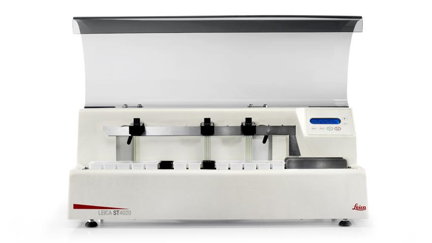
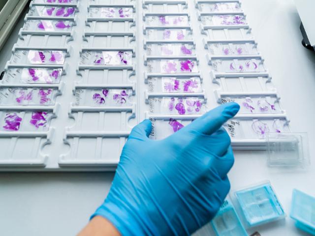
Leica ST4020 Small Linear Stainer


Overview
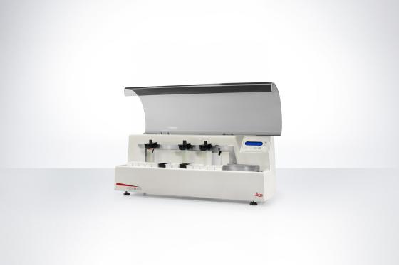
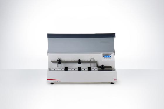
Leica ST4020 Small Linear Stainer delivers high-quality in a compact design.
The Leica ST4020 linear stainer combines the software programming features of a large linear stainer with a compact design. With advanced automation, the Leica ST4020 gives the user the freedom to start a run and dedicate their time to other urgent tasks meanwhile.
Flexible programming makes the Leica ST4020 ideal for many sample types. Turn to the Leica ST4020 when you need to run skin samples (Mohs specimens), frozen sections, fine needle aspirates and biopsies!
For in vitro diagnostic use.

Features
Save Money and Maintain Quality
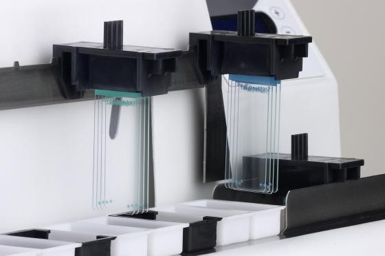
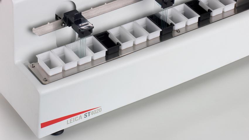
By using low reagent volumes, the Leica ST4020 linear stainer supports your laboratory to reduce waste and cut reagent costs. Optional agitation, running fresh water, and customizable protocols help to maintain a consistent diagnostic quality.
Accommodate Your Laboratory’s Workload
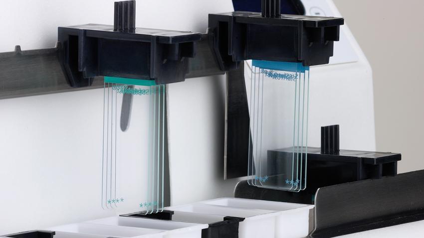
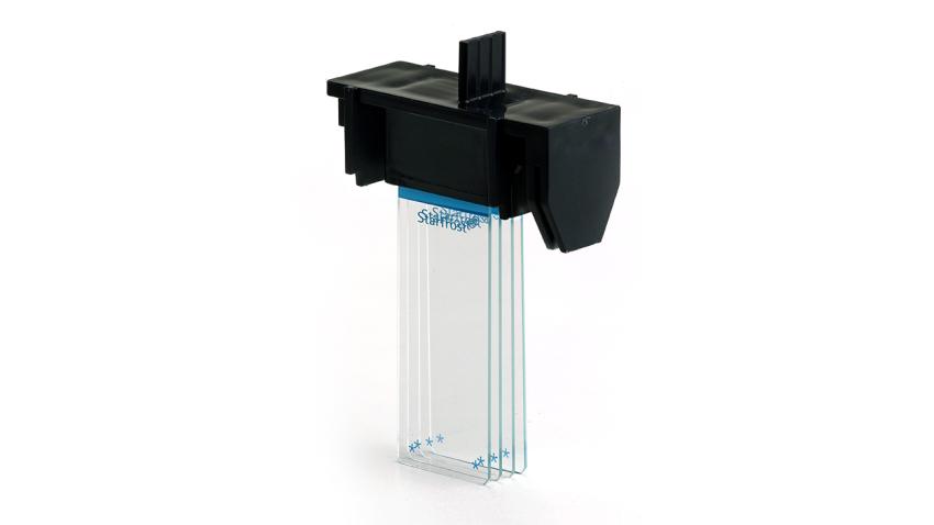
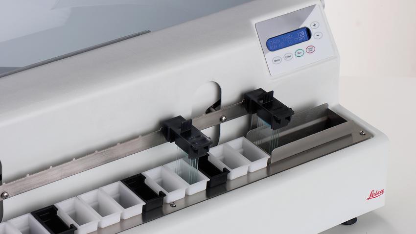
Multiple slides per run (1 to 4), a maximum throughput of more than 400 slides per hour and an exit tank for 16 slides help operators handle changing daily workloads.
Fits in Any Lab
Due to its compact size it is small enough to sit close to your cryostat to deal with even the most urgent samples.
Ensures Versatility and Freedom
The Leica ST4020 easily adapts to a wide range of laboratory workflows with flexible features including a variable protocol starting position, three optional running water stations and a programmable time per station of 2 to 300 seconds.
Think Lean and Secure
Accelerate turnaround with continuous loading and audible end-of-protocol notification.
Specifications
Downloads
Knowledge Hub
From tissue processing to Digital Pathology, our solutions empower labs with efficiency, precision, and reliability.
Though the H&E stain is a relatively simple stain to perform, there are a variety of artifacts that can interfere with a good stain. Artifacts can be attributed to a variety of causes.
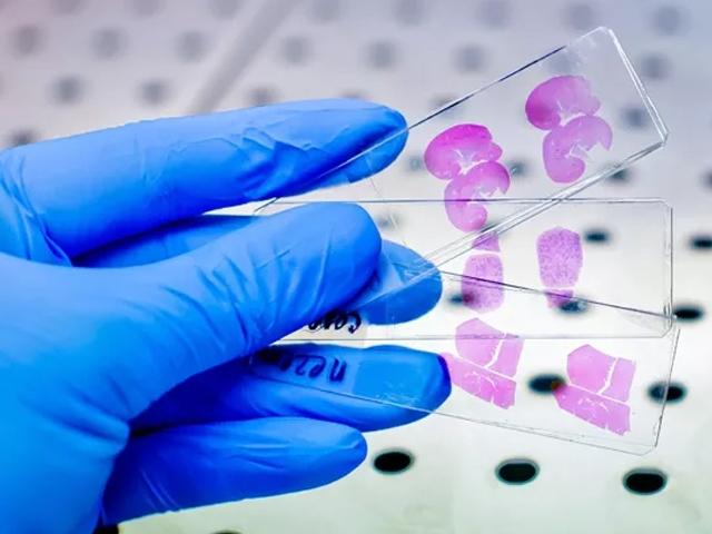
For routine diagnosis, the use of Hematoxylin and Eosin (H&E) is by far preferred for viewing cellular and tissue structure detail by pathologists.

Mucins are a part of a complex group called carbohydrates. Mucins are mucopolysaccharides; they are important in cell growth as they help regulate the flow of nutrients between capillaries and cells and are known as “The Glue of Life”.

Routine (or H&E) and special stains allow us to visualize otherwise transparent tissue under a microscope and are critical for tissue-based diagnosis.