News
Updates about Leica Biosystems.

Danaher has entered a partnership with AstraZeneca to scale precision medicine, including developing the next generation of AI-powered diagnostics This partnership aims to create a framework for rapid diagnostics research, development and commercialization Danaher, through its Centers for Enabling…

Melbourne, Australia – April 2, 2025 – Leica Biosystems, a global leader in anatomic pathology solutions, and Advanced Cell Diagnostics (ACD), a Bio-Techne brand and a global leader in spatial biology, today announced an expansion of their longstanding partnership to offer innovative spatial…
Suzhou, China - March 26, 2025 - Leica Biosystems, a global leader in anatomic pathology, today announced a strategic partnership with CellCarta, a globally recognized Contract Research Organization laboratory serving the biopharmaceutical industry. This partnership combines Leica Biosystems'…

January 28, 2025 – Nussloch, Germany. Leica Biosystems, a global leader in anatomic pathology, is known for its high-quality histology solutions. Today, the company announced the next iteration in its staining and coverslipping portfolio, the HistoCore CHROMAX Workstation (WS). Today, many…
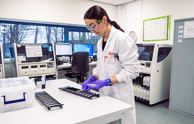
NEWCASTLE UPON TYNE, January 16, 2025 – Leica Biosystems, a global leader in anatomic pathology, has received a Clinical Laboratory Improvement Amendments (CLIA) certification for its state-of-the-art research and development facility in Newcastle, United Kingdom. A CLIA certification means a…