

")


Rápido y sencillo de utilizar para los técnicos de histología
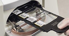
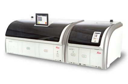
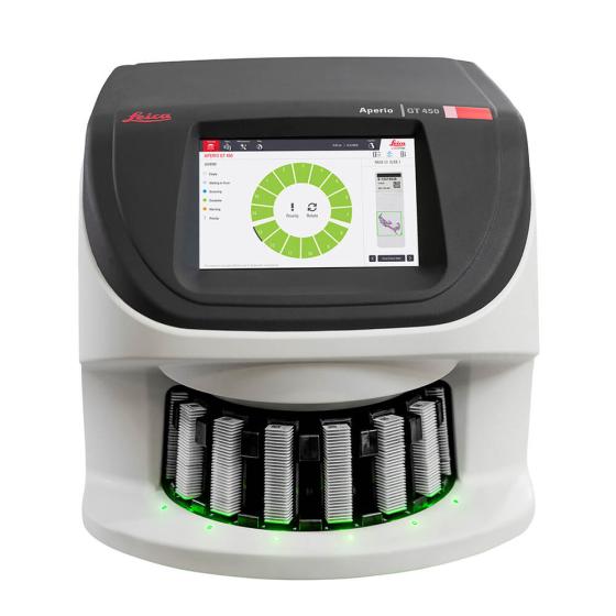
El escáner Aperio GT 450 es fácil de utilizar y ayuda a los técnicos histología a ofrecer resultados rápidos y fiables. Escaneado completo rápido y sencillo para que pueda centrarse en otras tareas del laboratorio: se pueden cargar 15 racks de 30 preparaciones (450 preparaciones en total) directamente desde el montador de cubreobjetos HistoCore SPECTRA CV en el Aperio GT 450. Se escanean automáticamente 81 preparaciones por hora a aumentos de 40x para un área de 15 mm x 15 mm.
- Carga continua de los racks sin contacto durante el escaneado
- Comprobación automática de la calidad de imagen durante cada escaneado para garantizar la calidad
- Asignar casos según su prioridad
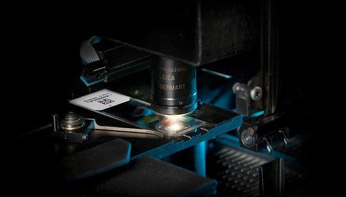
- Cada preparación se calibra con cada escaneado
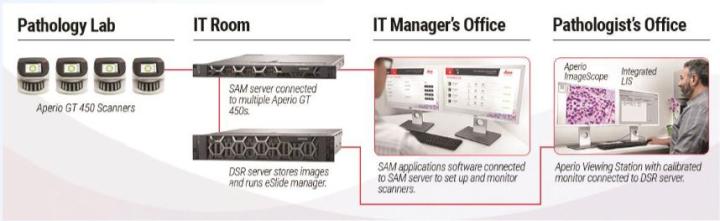
- La capacidad de 450 preparaciones y su diseño compacto y de reducidas dimensiones permite una fácil instalación en su laboratorio
- Detección de tejidos con una precisión del 99,5 % que puede ajustarse si es necesario*
- Tasa de éxito del escaneado del código de barras del 100 % al primer intento*
* Resultados de validación de VP-0450
“El Aperio GT 450 es muy fácil de usar y es un aliado en el laboratorio. Con sus altas velocidades de escaneado, comprobación automatizada de la calidad de imagen y escaneado prioritario de racks, me permite ser más productivo en mi jornada laboral”. - Asistente de laboratorio de Anatomía Patológica, NeoGenomics Laboratories


")


")