



Aperio iQC Software: AI-Powered Digital Pathology Quality Control



Updated Date: 7 October 2026
From Artifact to Accuracy, Intelligent Software Solutions for Faster, Reproducible Image QC
Summary
As digital pathology adoption accelerates, laboratories face growing pressure to maintain image quality while managing increasing slide volumes and limited technical resources. Although only a small percentage of whole slide images contain quality issues, every image typically requires review, creating a significant operational burden. Digital pathology demonstrates diagnostic concordance of approximately 98% with conventional microscopy while improving workflow efficiency and enabling scalable, networked pathology services.1–3 As adoption increases, ensuring the quality of whole slide images (WSIs) becomes critical to maintaining diagnostic confidence and operational performance.
Quality control (QC) remains a significant challenge because image artifacts such as missing tissue, focus issues, striping, pen marks, and air bubbles occur infrequently, typically affecting only 5-8% of slides. Yet laboratories spend as much as 25 full-time equivalent hours per day on manual QC activities, adding costs and stretching already limited technician resources. Research on the Low Prevalence Effect and vigilance decrement demonstrates that humans are inherently less effective at detecting rare events during repetitive, high-volume review tasks, making manual QC both resource-intensive and prone to inconsistency.4–5
Aperio iQC Software addresses this challenge through AI-powered quality control integrated directly into the Aperio GT scanner series workflow. Unlike manual review, which requires users to visually inspect every region of every WSI regardless of artifact likelihood, Aperio iQC Software automatically prioritizes images and regions most likely to contain quality issues. Trained and tested on more than 32,000 WSIs spanning diverse tissues, diseases, and staining protocols, the solution automatically detects six common digital and histological issues. Overall performance testing demonstrated >93% accuracy, sensitivity, and specificity, with performance exceeding 99% for selected artifact categories.
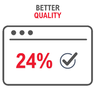
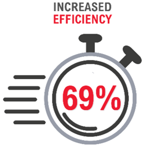
In a real-world evaluation conducted with the University of Heidelberg, Aperio iQC Software identified 24% more artifacts than manual review alone, reduced review time by 69% for digital pathology technicians, and increased slide review throughput by 2.5x. By automating QC and directing attention to the slides most likely to contain quality issues, Aperio iQC Software helps laboratories improve efficiency, standardize quality assessment, and strengthen confidence in digital pathology workflows.
Introduction
Digital pathology delivers significant workflow and diagnostic benefits, 6–8 but image quality issues remain a persistent challenge. Common whole slide image (WSI) artifacts—including missing tissue, clipped tissue, image striping, out-of-focus regions, pen marks, and air bubbles—can compromise image quality and disrupt downstream review. Today, identifying these issues often relies on manual inspection of every WSI, a time-consuming and subjective process that can be prone to inconsistency and human error. 1–2
Despite only 5–8% of slides typically exhibiting quality issues 3, laboratories must review every image to identify the small number that require attention. Real-world user data indicates that staff may spend up to 25 full-time equivalent hours per day performing quality control, with most users requiring 61–90 seconds to review a single WSI.3 This is an expensive, time-consuming body of monotonous work for laboratory technicians. There is a large body of research showing that humans are systematically poor at detecting rare events when reviewing large volumes of mostly normal cases. This is known as the Low Prevalence Effect and is directly relevant to pathology screening as well as other review tasks. 9–10 In addition, the vigilance decrement phenomenon, first coined by Mackworth et al. in 1948 demonstrates that performance declines over time when people continuously monitor for rare events.11 As such, quality control of large volumes of cases with low frequency of artifacts is intrinsically unsuitable for manual review and is an optimal low-risk, high-reward target to leverage AI in pathology.
To help address this challenge, Leica Biosystems developed Aperio iQC Software, an AI-powered quality control solution seamlessly integrated with the Aperio GT research scanners. Aperio iQC Software automatically detects and categorizes six of the most common digital and histological issues across a wide range of tissue types and stains. Slides are evaluated in an average of 31 seconds 4, with potential issues immediately highlighted on both the scanner console and centralized dashboard.
By automating image quality assessment, Aperio iQC Software standardizes the QC process, reduces manual review burden, and enables laboratory staff to focus on higher-value activities while helping ensure high-quality digital pathology images.
Software Application and AI Tool Development
To streamline quality control (QC) in digital pathology workflows, Leica Biosystems developed Aperio iQC Software, an AI-assisted application designed for seamless integration with the Aperio GT series of research scanners (Aperio GT 450, Aperio GT 180, and Aperio GT Elite). The software automatically detects and categorizes six common histological and digital issues—including missing or clipped tissue, image striping, out-of-focus regions, pen marks, and air bubbles—using AI algorithms developed specifically for whole slide image (WSI) quality assessment. 4–5
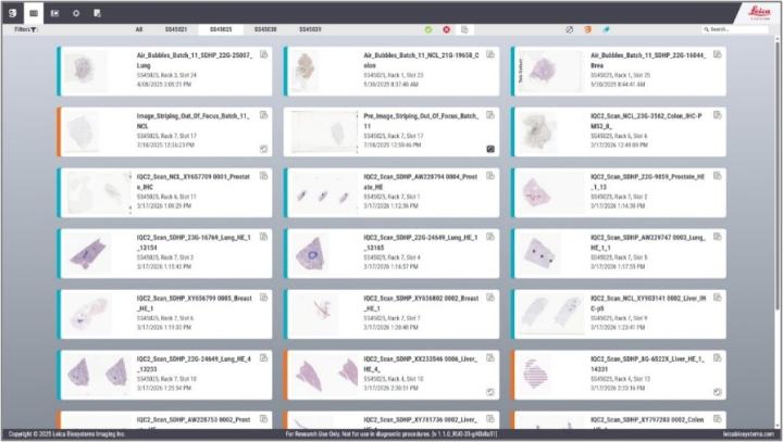
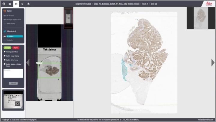
The Aperio iQC Software interface is designed to simplify QC review and accelerate issue identification. A centralized dashboard presents slides using a familiar slide-tray format and provides immediate visual indicators showing artifact status for each image. By directing users to slides and regions most likely to contain quality concerns, the software reduces manual review effort and helps users quickly focus on areas requiring attention.
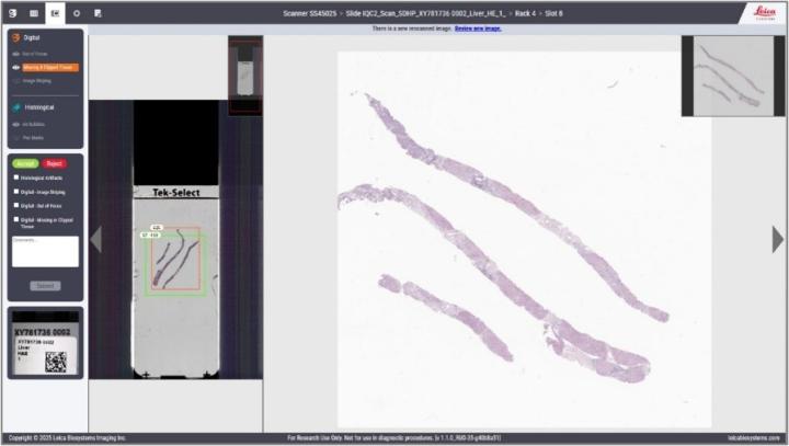
The viewing pane includes artifact-specific overlays that highlight the precise location of detected issues. For example, when an air bubble is detected, the corresponding tissue region is marked to allow immediate visualization and review without manually searching the entire slide.
Training and Testing Data Sets
Aperio iQC Software was trained and tested on a dataset of more than 32,000 whole slide images representing diverse sample types. Slides spanning 12 tissue types, H&E and IHC stains, benign and malignant disease states, and both biopsy and resection specimens were included in the dataset. This breadth and diversity are critical for developing AI models that generalize effectively across the wide variability encountered in routine practice. Ground truth was generated by a team of qualified annotators working alongside pathologists and laboratory experts using standardized quality-controlled annotation protocols. Combined with data from multiple scanner platforms, diverse tissue morphologies, and thousands of single- and multi-artifact examples, this breadth of training and testing data helps ensure robust performance and strong generalizability across real-world digital pathology workflows.
Quality, Trust, and Responsible AI
The Aperio iQC Software technology was developed under a comprehensive quality framework (ISO 13485) aligned with applicable internationally recognized standards for medical devices, software development, cybersecurity, and artificial intelligence. The research and clinical software versions adhere to numerous applicable ISO, IEC, IEEE, ANSI, and industry best practices that support product safety, reliability, security, and performance throughout the software lifecycle, as they pertain to in vitro diagnostic (IVD) and research products. 12–41 In practice, these quality processes include rigorous dataset governance, independent validation, risk-based software development, cybersecurity controls, and ongoing performance monitoring.
In addition, Aperio iQC Software technology follows emerging AI-focused standards and guidance for robustness, bias mitigation, data quality, and trustworthy AI, helping ensure consistent performance across diverse pathology workflows. Leica Biosystems quality management system is also aligned with ISO/IEC 42001, the international standard for AI management systems, reinforcing our commitment to the responsible development and deployment of AI-enabled solutions. By combining rigorous quality processes with internationally recognized standards, Aperio iQC Software delivers a standardized, reliable, and scalable approach to digital pathology quality control.
High Analytical Accuracy and Reliability
Overall performance testing demonstrated consistently strong performance across all targeted artifact types, with accuracy, sensitivity, and specificity exceeding 93% and reaching greater than 99% for some artifacts. For laboratories, this translates to dependable detection of image quality issues, fewer missed artifacts, and reduced time spent investigating false positives—helping drive both efficiency and confidence in digital pathology workflows.
Moreover, as the Aperio iQC Software artifact detection notifications are concurrently sent to both the software dashboard and the scanner console, quality control is embedded directly in the scanning workflow. This provides real time QC feedback to laboratory staff enabling rapid identification of issues while the slide is still on the scanner, thus optimizing the process and delivering QC information where and when it is needed.
Real-World Performance
In a study of 200 prostate whole slide images, conducted with the University of Heidelberg, both a digital pathology technician and a pathologist identified substantially more artifacts when using Aperio iQC compared with manual review alone. The Aperio iQC Software detected 24% more artifacts, which were not identified by the pathology technician during manual review. These results demonstrate that AI-assisted quality control can help uncover image quality issues that may otherwise be missed, supporting more consistent and reliable slide assessment. 4–5
Beyond improving artifact detection, Aperio iQC Software significantly reduced review times with 69% faster review by the digital pathology technician compared to manual review. Overall, Aperio iQC Software increased whole slide image review throughput by 2.5×, enabling laboratories to review more slides in less time while maintaining confidence in image quality.
Conclusions
Aperio iQC Software combines cutting-edge, AI-powered artifact detection with workflow automation to help laboratories improve digital pathology quality control. Trained and tested on a diverse dataset spanning tissue types, disease states, and staining protocols, the solution demonstrated high accuracy, sensitivity, and specificity across all targeted artifact categories.
In real-world evaluations, Aperio iQC Software enabled users to identify 24% more image quality issues than manual review alone while significantly reducing review time by 69%. This reduction in manual QC time can be directly recognized in labor cost savings, while indirect benefits include reduction in monotonous laboratory tasks for pathology technicians and standardization of quality reviews. By automatically screening slides and highlighting potential artifacts, the software helps laboratories focus attention where it is needed most.
The result is a more efficient and standardized quality control process—reducing manual review burden, increasing slide review throughput by up to 2.5×, and supporting confidence in the quality of whole slide images. By transforming QC from a subjective, labor-intensive task into an automated workflow, Aperio iQC Software allows pathology teams to spend less time searching for issues and more time on higher-value activities.
The Aperio iQC Software technology is available in different formats in different geographies. Please check with your local representative for details of availability.
Aperio iQC Software is For Research Use Only. Not for Use in Diagnostic Procedures.
In the USA Aperio iQC DX Software (K253561) is FDA 510(K)-cleared For In Vitro Diagnostic Use.
Aperio iQC DX Software is IVDR CE-marked For In Vitro Diagnostic Use.
About the presenters
Sheheryar Arshad, PhD, is the Technical Lead for Deep Learning and Computer Vision Science at Leica Biosystems. He specializes in computer vision and scalable whole-slide image analysis, advancing AI-driven solutions for digital pathology. His work on automated artifact detection and image quality is anchored in improving workflow efficiency and diagnostic confidence. Dr. Arshad holds a PhD in Computer Engineering with focus on AI from the University of Texas and has authored 17+ publications.
Dr. Colgan has over a decade of experience in the digital pathology sector and is focused on how this new and disruptive technology can be leveraged to provide real benefits in both the healthcare and research domains. Prior to working with Leica Biosystems, she came from a research background with a BSc in Biotechnology and a PhD in Vascular Biology from Dublin City University, Ireland.
Dr. Catherine Conway is Director of Clinical AI Applications for Digital Pathology at Leica Biosystems. During her career, Catherine has led high-impact scientific and technological initiatives across academia, medical device companies, and innovative startups to advance both research and clinical practice. Her career is also distinguished by a focus on bridging scientific rigor with practical, real-world application, and includes extensive work in highly regulated environments, augmented with a strong publication record. She holds a BSc and PhD in Cancer Research and completed a Postdoctoral fellowship at the National Cancer Institute (NCI), National Institutes of Health (NIH), USA. She is deeply committed to translating science into solutions that make a tangible difference in patient outcomes and medical progress.
Referencias
[1] Farahani N, Parwani AV, Pantanowitz L. Whole slide imaging in pathology: Advantages, limitations, and emerging perspectives. Pathology and Laboratory Medicine International. 2015;7:23-33.
[2] Hanna MG, Reuter VE, Hameed MR, Tan LK, Chiang S, Sigel C, Hollmann T, Giri D, Samboy J, Moradel C, Rosado A, Otilano JR III, England C, Corsale L, Stamelos E, Yagi Y, Schüffler PJ, Fuchs T, Klimstra DS, Sirintrapun SJ. Whole slide imaging equivalency and quality considerations in digital pathology. Modern Pathology. 2019;32(7):916-928.
[3] Leica Biosystems. Voice of Customer Survey. Internal customer feedback survey. Leica Biosystems; 2025.
[4] Balakrishnan BTS, Legnar M, Kaib P, Weis CA, Valand J, Wang J, Eslami Manoochehri H, Arshad SA, Khorramshahi P, Slocomb J, Baty A, Clardy W, Sutcu AE, Ferber D, Choksi L, Miller S, Colgan O, Conway C. Aperio iQC Software for automated quality control in digital pathology. Poster presented at: 12th Digital Pathology & AI Congress Europe; 2025; Heidelberg, Germany.
[5] Wang J, Eslami Manoochehri H, Baty A, Valand J, Clardy W, Arshad SA, Khorramshahi P, Slocomb J, Miller S, Conway C. Aperio iQC Software: AI-driven artifact detection for quality control in digital pathology. Poster presentation. Leica Biosystems; 2026.
[6] Azam AS, Miligy IM, Kimani PKU, Maqbool H, Hewitt K, Rajpoot NM, Snead DRJ. Diagnostic concordance and discordance in digital pathology: A systematic review and meta-analysis. Journal of Clinical Pathology. 2021;74(7):448-455.
[7] Pantanowitz L, Sinard JH, Henricks WH, Fatheree LA, Carter AB, Contis L, Beckwith BA, Evans AJ, Lal A, Parwani AV; College of American Pathologists Pathology and Laboratory Quality Center. Validating whole slide imaging for diagnostic purposes in pathology: Guideline from the College of American Pathologists Pathology and Laboratory Quality Center. Archives of Pathology & Laboratory Medicine. 2013;137(12):1710-1722.
[8] Iwuajoku V, Ekici K, Haas A, et al. An equivalency and efficiency study for one year of digital pathology for clinical routine diagnostics in an accredited tertiary academic center. Virchows Archiv. 2023;483:583-592.
[9] Wolfe JM, Horowitz TS, Kenner NM. Rare items often missed in visual searches. Nature. 2005;435(7041):439-440. doi:10.1038/435439a
[10] Wolfe JM, Horowitz TS, Van Wert MJ, Kenner NM, Place SS, Kibbi N. Low target prevalence is a stubborn source of errors in visual search tasks. Journal of Experimental Psychology: General. 2007;136(4):623-638. doi:10.1037/0096-3445.136.4.623
[11] Mackworth NH. The breakdown of vigilance during prolonged visual search. Quarterly Journal of Experimental Psychology. 1948;1(1):6-21. doi:10.1080/17470214808416738
[12] ISO 13485. Medical devices – Quality management systems – Requirements for regulatory purposes.
[13] ISO 14971. Medical devices – Application of risk management to medical devices.
[14] ISO 15223-1. Medical devices – Symbols to be used with medical device labels, labelling and information to be supplied – Part 1: General requirements.
[15] EN ISO 18113-1. In vitro diagnostic medical devices – Information supplied by the manufacturer (labelling) – Part 1: Terms, definitions and general requirements.
[16] EN ISO 18113-3. In vitro diagnostic medical devices – Information supplied by the manufacturer (labelling) – Part 3: In vitro diagnostic instruments for professional use.
[17] ISO 7000/IEC 60417. Graphical symbols for use on equipment.
[18] BS EN ISO 20417. Medical devices – Information to be supplied by the manufacturer.
[19] IEC 62304. Medical device software – Software life cycle processes.
[20] ISO/IEC 27001. Information technology – Security techniques – Information security management systems – Requirements.
[21] IEC TR 80001-2-2. Application of risk management for IT networks incorporating medical devices – Part 2-2: Guidance for the disclosure and communication of medical device security needs, risks and controls.
[22] NIST SP 800-53. Security and Privacy Controls for Information Systems and Organizations.
[23] IEC TS 62443-1-1. Industrial communication networks – Network and system security – Part 1-1: Terminology, concepts and models.
[24] IEC 62366-1. Medical devices – Part 1: Application of usability engineering to medical devices.
[25] ANSI/AAMI HE75. Human factors engineering – Design of medical devices.
[26] ISO 17050-1. Conformity assessment – Supplier's declaration of conformity – Part 1: General requirements.
[27] ISO/IEC 8183. Information technology – Artificial intelligence – Data life cycle framework.
[28] ISO/IEC TR 24028. Information technology – Artificial intelligence – Overview of trustworthiness in artificial intelligence.
[29] ISO/IEC 24029-2. Artificial intelligence – Assessment of the robustness of neural networks – Part 2: Methodology for the use of formal methods.
[30] IEEE 2801. IEEE Recommended Practice for the Quality Management of Datasets for Medical Artificial Intelligence.
[31] ISO/IEC 42001. Information technology – Artificial intelligence – Management system.
[32] DIN SPEC 13288. Guideline for the development of deep learning image recognition systems in medicine.
[33] ISO/IEC TR 24027. Information technology – Artificial intelligence – Bias in AI systems and AI-aided decision making.
[34] ISO/IEC TR 24029-1. Artificial intelligence – Assessment of the robustness of neural networks – Part 1: Overview.
[35] AAMI CR34971. Guidance on the application of ISO 14971 to artificial intelligence and machine learning.
[36] ISO/IEC 4213. Information technology – Artificial intelligence – Assessment of machine learning classification performance.
[37] ISO/IEC TR 24372. Information technology – Artificial intelligence – Overview of computational approaches for AI systems.
[38] ISO/TR 24291. Health informatics – Applications of machine learning technologies in imaging and other medical applications.
[39] ANSI/CTA 2090. The Use of Artificial Intelligence in Health Care: Trustworthiness.
[40] ISO/IEC 5259-1. Artificial intelligence – Data quality for analytics and machine learning (ML) – Part 1: Overview, terminology and examples.
[41] IEEE 2841. IEEE Recommended Practice for Framework and Process for Deep Learning Evaluation.
Related Content


El contenido de Leica Biosystems Knowledge Pathway está sujeto a las condiciones de uso del sitio web de Leica Biosystems, disponibles en: Aviso legal.. El contenido, incluidos los webinars o seminarios web, los recursos de formación y los materiales relacionados, está destinado a proporcionar información general sobre temas concretos de interés para los profesionales de la salud y no está destinado a ser, ni debe interpretarse como asesoramiento médico, normativo o jurídico. Los puntos de vista y opiniones expresados en cualquier contenido de terceros reflejan los puntos de vista y opiniones personales de los ponentes/autores y no representan ni reflejan necesariamente los puntos de vista ni opiniones de Leica Biosystems, sus empleados o sus agentes. Cualquier enlace incluido en el contenido que proporcione acceso a recursos o contenido de terceros se proporciona únicamente por comodidad.
Para el uso de cualquier producto, debe consultarse la documentación correspondiente del producto, incluidas las guías de información, los prospectos y los manuales de funcionamiento.
Copyright © 2026 Leica Biosystems division of Leica Microsystems, Inc. and its Leica Biosystems affiliates. All rights reserved. LEICA and the Leica Logo are registered trademarks of Leica Microsystems IR GmbH.