15 result(s) for 'Colon'
1 - 10 of 15 results for 'Colon'
Sort by
Show
Slide 1
Welcome
Slide 2
Slide 3
Discuss Pre-Analytics and the impact of improper fixation and artifacts on tissue processing
Discuss Fixation and the impact of incomplete fixation on tissue processing
Describe the impact of improper Prosection
Determine satisfactory Processing of samples
Explain why routine Maintenance is a critical success factor to proper tissue processing
Slide 4
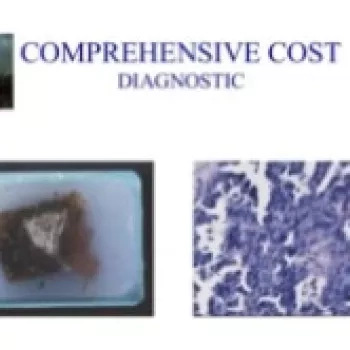
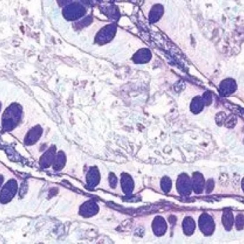
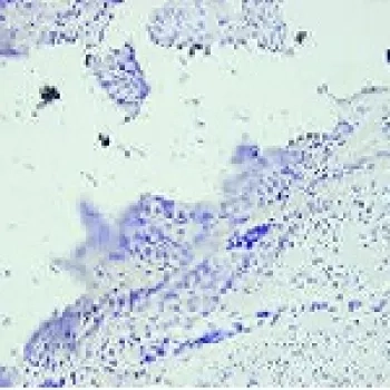
This slide has two examples of what the ideal sections in a perfect world look like under the scope. The skin section on the left is clean looking with the Eosin staining the components of the dermis with different shades. The colon section on the right is crisp with well-defined nuclei and cilia. There is no background staining or muddiness to the stain.
Slide 5
One artifact seen the lab that is n...
One of the most fundamentally critical elements of diagnostic histopathology is first the ability to suspend all cellular activity in tissue and prevent degradation, and secondly to process that specimen in a manner that facilitates subsequent steps such as...
Slide 1
Hello, hello; thank you so much for that introduction. Let's get started. Oh, I'm sorry, I just need to hit this next button here. So my name is Mark Lawson.
Thank you for joining us in this webinar. I'm here to present BOND RX tips, tricks, and optimization. It's going to be a user guide from the BOND RX and chromogenic multiplexing in the research application.
Slide 2
So, my name is Mark Lawson. I'm an application specialist on the Life Sciences team at Leica Biosystems I provide technical support for the Life Sciences portfolio, including but not limited to the BOND RX, the BOND RXm, and a wide array of reagents. So I've worked in the histology field for about 15 years and in both clinical and research spaces. I started off as a histotechnologist and worked my way up...
The content of this webinar will encompass detailed technical descriptions of novel and state of the art methods to decalcify tissue samples that will be embedded in paraffin or for frozen sectioning for molecular and nucleic acid analysis for basic research...
Biomarkers
AE1/AE3 (1)
AMACR (2)
BCL6 (1)
CD163 (2)
CD20 (2)
CD3 (9)
CD4 (5)
CD68 (5)
CD8 (10)
CDX2 (2)
CK20 (5)
Ck5 (4)
...
Special stains that are used for the evaluation of mucins, mucin-like molecules and other carbohydrate containing macromolecules remain in demand and are utilized frequently in the histology laboratory.
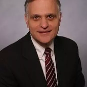
Dr. Steve A. Kargas is the current Director, Surgical Pathology at CSI Laboratories, Atlanta, GA. He received his medical degree and post doctorate at University of Wisconsin-Madison and his AP residency at Stanford and CP residency at Cedars Sinai Medical Center, LA., CA. Dr. Kargas is highly involved in improving specializing in evaluation, diagnostic immunohistochemical analysis and molecular testing of solid tumors including carcinomas of unknown primary site, soft tissue, colon, prostate, breast, prognostic/diagnostic cases, MMRP, MSI PCR testing, hepatocellular carcinoma, lung including small cell carcinoma vs. non-small carcinoma, and providing solutions to anatomic pathology needs of clients. He is a member of CAP, USCAP, ASCP, ASQ, and ASC.
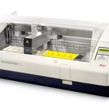
The proven Leica ST5010 Autostainer XL provides reproducible, consistent high-quality staining, and increased workload throughput compared to manual staining.
Combine it with the CV5030 Glass Coverslipper to create a workstation that eliminates the manual handling of slide racks between staining and coverslipping.
To give the user flexibility, choose between your preferred consumables manufacturer or validated Leica Biosystems consumables and protocols which provide superb staining and coverslipping quality.
Get tips for better ISH staining in this guide. Each step provides a valuable reminder of good histology practice and also helps with troubleshooting when unacceptable results do occur.