
Tips & Tricks to Multiplexing: How to Choose Chromogen Colors for Multiplex and Detection Systems for Multiplex Assays

Multiplexing is an important tool for simultaneous detection of multiple markers within a single tissue section.
Watch our 2 short videos below to learn more about multiplexing and download the complementary infographics to keep the points on hand when you need to revisit the information!
How to choose chromogen colors for multiplex
If you have opted for chromogenic multiplex staining, there are additional points to consider: the choice of colors and the order of color application. While this video won’t tell you exactly what, when, and where to use different chromogens, it will provide you with some tips to consider.
Check out below and learn with Larry from Leica Biosystems about the tips to consider for chromogenic multiplex staining into your tissue-based research.
10 Tips for Chromogenic Multiplexing
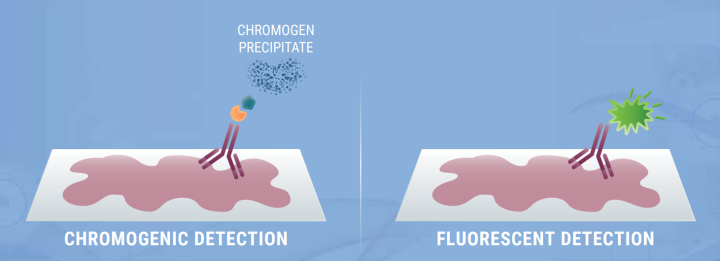
Chromogenic multiplex staining needs careful planning and consideration to produce a multi-color, multi-target, permanent slide that is resistant to photo-bleaching, and perfect for brightfield imaging and downstream analysis.
Consider using lighter chromogen colors
- Lighter chromogen colors may be easier on the eyes to visualize especially when multiple chromogens are involved.
Optimize the chromogen sequence
- DAB can overstain and occlude previously stained sites. Consider its suitable place in the assay.
Consider your choice of preferred counterstain
- Ensure the counterstain provides appropriate contrast and does not interfere with chromogen interpretation.
Consider the compatibility of your chromogens
- Consider the compatibility of your chosen chromogens with your required mounting media and preferred dehydration methods.
Consider the order of your markers
- Determine which antigens are robust or susceptible to degradation following multiple rounds of antigen retrieval
Test the stability of different chromogens
- Use chromogens with signals that remain strong earlier in the assay, and less robust signals later in the experiment.
Consider selecting the chromogen colors
- Consider selecting the chromogen colors for spatially close targets first and then build the assay further.
Consider the rate of chromogen precipitation
- Faster precipitating chromogens may be better suited for low expressed proteins. Consider slower precipitating chromogens for highly expressed proteins.
Consider high vs low expression
- Consider using a strong color chromogen for low expression, or low in quantity markers. Followed with a weaker color chromogen for high expression, or high in quantity markers.
Consider color combinations
- For colocalized markers, aim to use chromogens that will create a third color when combined.
Detection systems for multiplex assays
There are many decisions that must be made when adopting multiplex staining, such as choosing chromogenic or immunofluorescent multiplex assays, as well as the order and colors of chromogens and fluorophores. However, the decisions don’t stop here as there are different detection systems available for multiplex assays that can increase sensitivity and enhance the signal-to-noise ratio when detecting markers of interest. We will cover a few of these examples in this video.
Unsure of what detection systems are available for multiplex assays? Check out the infographic below that showcases some examples of detection systems available for multiplex assays that can increase sensitivity and enhance the signal-to-noise ratio when detecting markers of interest.
Multiplex Detection Systems
There are several detection systems available to visualize multiplex staining that are: direct primary antibody detection, or indirect detection with ABC, polymer-linked antibodies, with, or without tyramide amplification, and finally, oligonucleotide labelled primary antibodies with complementary probes. Whichever detection system you decide to use in your multiplex assay, optimization is vital.
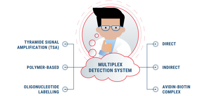
Direct Detection
- Suitable for highly expressed proteins
- Can avoid cross-reactivity by species-specific secondary antibodies
Indirect Detection
Tyramide Signal Amplification Detection System

Don't Miss Out! Subscribe To Research Link Today To Get Quaterly Updates On Our Latest Life Science Endeavours
For research use only. Not for use in diagnostic procedures.
About the presenter
Rhian is a Scientist from Swansea University in Medical and Healthcare Studies and was featured in several collaborative publications. Rhian’s research-based background focused on tissue-based pathology in Multiple Sclerosis, primarily using immunohistochemical analysis and in vitro molecular techniques. She spent a short period conducting routine PCR testing for COVID-19 at the end of 2020.
Related Content


Leica Biosystems content is subject to the Leica Biosystems website terms of use, available at: Legal Notice. The content, including webinars, training presentations and related materials is intended to provide general information regarding particular subjects of interest to health care professionals and is not intended to be, and should not be construed as, medical, regulatory or legal advice. The views and opinions expressed in any third-party content reflect the personal views and opinions of the speaker(s)/author(s) and do not necessarily represent or reflect the views or opinions of Leica Biosystems, its employees or agents. Any links contained in the content which provides access to third party resources or content is provided for convenience only.
For the use of any product, the applicable product documentation, including information guides, inserts and operation manuals should be consulted.
Copyright © 2026 Leica Biosystems division of Leica Microsystems, Inc. and its Leica Biosystems affiliates. All rights reserved. LEICA and the Leica Logo are registered trademarks of Leica Microsystems IR GmbH.