Automated Multiplex Immunofluorescence


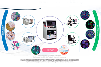
Cancer research continues to push the boundaries with new advancements in tissue analysis and biomarker detection. Now, more than ever, there is significant emphasis on understanding the underlying interaction between the immune system and the tumor microenvironment. Multiplexing immunofluorescence (mIF) has greatly increased our understanding of solid tumor biology and immunology, including tumor-infiltrating lymphocytes and cancer-induced architectural alterations, and aided in novel immunology discoveries. In this webinar, we will discuss how Akoya Biosciences Phenoptics assays support quantitative mIF to overcome the limitations imposed by conventional IHC methodologies. We will also discuss how our Opal assay kits and reagents can be integrated with the Leica BOND RX to automate your staining workflow to support consistent results for high-throughput studies.
Learning Objectives
- Understand the limitations of conventional IHC , and the benefits of transitioning to multiplex immunofluorescence
- Learn how to design, optimize and analyze your multiplex assay panel
- Understand how the BOND RX enables innovations like the Opal assay
Webinar Transcription
Hello everyone and welcome to today's live webinar automated multiplex immunofluorescence presented by Dr. Bethany Remeniuk and Traci DeGeer. I'm Christy with Labroots and I'll be your moderator for today's event. Today's educational web seminar is presented by Labroots and brought to you by Leica Biosystems. For more information on our sponsor, please visit the sponsor tab at the left side of your screen and click on the logos to be directed to the sponsor website.
Before we begin, I would like to remind everyone that this event is interactive and we encourage you to participate by submitting as many questions as you want at anytime you want during the presentation. To do so, simply type them into the ask a question box and click send. We will answer as many questions as we have time for at the end of the presentation. If you have trouble seeing or hearing the presentation, click on the support tab found at the top right of the presentation window. Or use the ask a question box to let us know you're experiencing a problem.
This presentation is educational and thus offers continuing education credits. Please click on the continuing education credits tab located at the top right of your presentation window and follow the process to obtain your credits. I would now like to present today's speakers doctor Bethany Remeniuk, Global Application Scientist, Akoya Biosciences, and Tracy DeGeer, Director, Advanced Staining Innovation, Leica Biosystems. For complete biographies on our speakers, please visit the biography tab at the top of your screen. Bethany and Tracy, you may now begin your presentation.
Thank you, Christy for that introduction. Hello everyone. My name is Bethany Remeniuk and I am the Global Application Scientist at Akoya Biosciences. Today, we will be discussing the advantages of utilizing Akoya's opal multiplex immunohistochemistry, automated detection kits on the Leica Biosystems BOND RX auto stainer to meet your research needs. In addition, I will be highlighting some tips and recommendations to help design your multiplex panel and optimize your staining. However, before we can delve into this, we must first take a step back to understand how these two platforms combined help to facilitate a streamlined workflow and drive cancer research forward.
From 1991 to 2015, cancer deaths in the United States alone fell by 26%. This is due in part to significant advances in the treatment of cancer patients, including the use of more personalized therapies for individuals as well as FDA-approved immunotherapy treatments such as Anti-PD-1 and Anti-PD-L1. However not all individuals who test positive for these markers will respond to targeted treatment.
The Underlying Biology of the Tumor Microenvironment is Complex
To help understand why we may have responders and non-responders, we must first acknowledge that the biology and composition of the tumor microenvironment is very complex. There are several specialized cell types, including immune mediated cells such as macrophages and B&T cell lymphocytes that interact directly and indirectly with tumor cells, resulting in an ever-evolving relationship between immune cells trying to destroy the tumor cells while simultaneously the tumor cells attempting to evade and promote their own self growth.
Because of this, there are several influencing factors that need to be taken into consideration when interrogating the tumor microenvironment, including cellular phenotypes that could influence the biology of the tumor, the functional status of the surrounding cell types, as well as other contributing factors such as inflammatory mediators and how these all interact within the spatial biology of the tumor microenvironment. Having a greater understanding of the relationships happening within the tumor microenvironment are key to improving diagnosis and designing new therapeutic regimens.
Limitations of Current Analysis Platforms
Currently, clinical laboratory tests are automated and performed by calibrated machines to reduce human error. However, most anatomic pathology and disease diagnosis is still based upon physician interpretation of microscopic tissue characteristics such as cells, markers, and tissue architecture. In fact, this visual assessment using conventional immunohistochemistry remains the gold standard for cancer diagnosis.
Despite this, it is recognized that visual assessment can be subjective and interpretation of results can differ between pathologists. Additionally, because immunohistochemistry such as DAB can only be conducted for a single marker on tissue samples, very little information can be obtained about the spatial context and interplay of other cellular phenotypes within the tumor microenvironment.
On the other hand, genomics and proteomics studies including flow cytometry and DNA microarrays, provide significant quantitative information regarding different cellular phenotypes within the tumor. However, the spatial context is lost as analysis needs to be conducted using homogenized tissue and thus were at a crossroads. Where on one platform we are able to retain morphology but lose phenotypic information or conversely, have a wealth of phenotype data but lose spatial biology and interactions between them.
Immunofluorescence Reveals Complex Biology
To advance research forward, we need to be able to retain the spatial biology of the tumor microenvironment while simultaneously identifying and labeling multiple markers of interest. This is where immunofluorescence is truly able to bridge the divide. Immunofluorescence overcomes the limitations of conventional immunohistochemistry, which are constrained by detecting two or three markers of interest, by allowing for multiplexing of several markers. By using this methodology, researchers can conserve their tissue samples, while identifying and characterizing cellular interactions.
Immunofluorescence can then identify and reveal weak expressors that may be lost using conventional immunohistochemistry staining, and furthermore, the balancing of signals can allow researchers to detect multiple markers that colocalize on the same cell. Lastly, immunofluorescence can provide better signal for quantification and data analysis.
Challenges with Conventional Immunofluorescence
While there are many benefits associated with immunofluorescence, there are challenges with conventional methodologies that can unfortunately limit your staining success. First, staining will need to be done using antibodies raised in different species, as secondary antibodies may crossreact and ruin your results. This unfortunately can be restricting as some rare antibodies may only be raised in one species.
Next, some antigens may be sheltered from the primary antibody due to stearic hinderance. Furthermore, signals can be unbalanced or low if you have an overabundance of a marker or very little expression of another. Other factors that may impact your results include background staining, contributing tissue autofluorescence, which can be a result of the tissue itself as well as how the tissue was processed, and photobleaching, or quenching, of the fluorophores as they are exposed to light.
Opal Fluorophores for Multiplexing
Thus, the Opal flurophores were created to overcome the limitations of conventional immunofluorescence. As part of the synoptics workflow, Opal dyes are accessible to anyone who has previously worked with standard immunohistochemistry. Opal allows for detection of multiple cellular phenotypes to be visualized and quantified simultaneously in the same formalin-fixed paraffin embedded tissue sections, enabling researchers to study the complex relationships and distribution of these cells while retaining spatial context within the tumor microenvironment.
With Opal you can select any primary antibody you wish for immunohistochemistry detection based upon their performance, rather than being relegated to a species type. And lastly, Opal's multiplex assays and kits provide a practical workflow that allows for the simultaneous detection of upwards of eight biomarkers of interest post tapping nuclear counter stain within a single tissue section.
Automated Staining with Opal on the BOND RX
Akoya offers Opal automated IHC detection kits for researchers to perform Opal multiplex staining on one of the leading research automated stating platforms, the BOND RX by Leica Biosystems. Automation provides you with the flexibility to support the dynamic demands of translational research. Open platform promotes a simplified workflow that frees up users’ time for other research endeavors.
High-throughput Opal BOND protocols can perform upwards of seven color immunofluorescence staining on 30 slides, which can be performed overnight as opposed to typically running manually, which may take two to three days. And lastly, automation can achieve quality, consistency and reproducibility with every sample and supports the transition into clinical studies.
How to Stain with Opal on the BOND RX
How does one go about staining with Opal using the BOND RX? As was previously mentioned, Opal dyes allow for the use of any standard unlabeled primary antibody, including multiple antibodies raised in the same species. Here we have a microscope slide that has been barcoded to recognize to be recognized by the BOND RX. After introduction of the primary antibody, the Opal polymer, HRP, is applied.
The Opal system then uses tyramide signal amplification, or TSA, to amplify IHC detection by covalently depositing multiple fluorophores near the targeted antigen. After labeling is complete, antibodies are removed in a manner that does not disrupt the Opal fluorescence signal, allowing for the next target to be detected without antibody cross reactivity. These steps are repeated for all markers of interest. The Opal technology thus enables development of multiplex assays with balanced quantitative signal for both rare and abundant targets of interest.
Opal Dyes are Stable Through ER Repetition in the BOND RX
Moving forward, we wanted to test the stability and reproducibility of Opal on the BOND RX. First, to confirm fluorescent signal stability of the Opal dyes on the BOND RX, we conducted a series of repeated stripping steps with each Opal fluorophore and determined that over the course of six repeated high intensity epitope retrievals that the Opal dye signal remains stable for all fluorophores, including our classic six fluorophores and our latest edition of fluorophores including the Opal 480.
Detect Co-localized Markers with Opal
Next, to demonstrate Opal's ability to detect markers in the same cellular compartments without interference from TSA deposition, we stained tonsil with both CD3 and CD8, as they are often found in close proximity with one another on the same cell. On the left we have a merged overlay of the CD3 and CD8 channels where signal overlap appears in white. Then we have the individual channels appearing to the right. As you can see, TSA does not interfere with the detection of co-localization of markers of interest.
BOND RX Produces Consistent Opal Staining: Monoplex
Next, we wanted to test the consistency and reproducibility of the BOND RX at staining Opal. Here we ran monoplexes staining for CD20 using Opal 520 on serial sections of tonsil for all 30 slide positions. We demonstrate that slide variability between all 30 slides for per cell expression of CD20 produced a coefficient of variation less than 5%, indicating that the BOND can produce consistent and reliable staining results.
BOND RX Produces Consistent Opal Staining: Multiplex
For the next testing procedure, we assessed the BOND RX’s ability to reliably stain 15 tonsil serial sections for seven color multiplex panels. For each of the Opal fluorophores, we analyzed the mean counts of the top 20 brightest cells per slide and found that for each marker there was a calculated CV of 14% or less. Which, given the nature of serial sections, speaks to the consistency of both Opal staining as well as the functionality and reliability of the BOND RX in producing consistent results for all your staining needs.
Opal Workflow Overview on the BOND RX
Next, we're going to delve into some tips, tricks, and recommendations to help optimize your Opal staining. Here on the left we have an overview of how staining is run on the BOND RX using the newest Opal Polaris immunohistochemistry automated detection kit, which includes our newest fluorophores, the Opal Polaris 480, and the Opal Polaris 780.
Opal protocols are auto populated on the BOND RX and can be used as they are for all of our other kits that we have. However, if you wish to do staining with the Opal Polaris kit, you will need to modify that protocol to include Opal Polaris 780 at the end of the staining run. At the end of this presentation as well, I will also share some additional staining resources to help support your staining endeavors.
Considerations for Monoplex Development
First we have some recommendations from Akoya to help in your monoplex development. First, we recommend that you use tonsil to optimize all of your monoplexes. This is ideal because tonsil has several markers of interest that are highly expressed, making it ideal to help optimize your primary antibody dilution.
We recommend going ahead with a series of three DAB titrations and then optimizing from there based on staining pattern and intensity. Once you feel you've optimized your primary antibody dilution using DAB, we recommend then converting it to immunofluorescence. Here then you would want to pick an ideal Opal pairing to go with the primary antibody and some factors to consider include co-expression. If you happen to be looking at multiple markers of interest on the same cellular compartment, you might want to choose an Opal pairing for that primary antibody that is perhaps a little bit far removed from the other marker of interest that you are looking to image.
For example, you may want to choose Opal 520 paired to CD3 and then maybe Opal 570 or Opal 620 to look at CD8. Furthermore, you also want to take into consideration whether you are investigating rare versus abundant markers. If you are looking at markers that may not be highly expressed or weak expresses, you may want to pair them with an Opal that is very bright. In this case, Opal Polaris 480 or Opal 520 versus if you happen to have a blended marker of interest such as cytokeratin, you may want to pair that with some of the more lower bright Opal dyes, including Opal 690 and Opal Polaris 780.
For Opal concentrations, we recommend starting at a 1 to 150 dilution and then titrating up or down as needed. We like you to remain within already predetermined primary antibody dilution.
Next, you'll want to go ahead and assess your target brightness, and we recommend that target brightness counts fall within a range of 10 to 30, except for Opal Polaris 780 because it is near the infrared. We recommend that you target anywhere from a range of 1 to 10. This can be done using our one platform, which is free, it's Phenochart. You can go ahead and then look at the normalized counts and then you can adjust your Opal titration from there.
Once you get done optimizing your tonsil monoplexes, we recommend transitioning those dilution factors onto project tissue samples and reoptimizing as needed. We recommend not really adjusting the primary and body dilution factor and trying to titrate out your Opal.
Considerations for Library Development
Next, for each project, Akoya recommends that you create a new library slide set to ensure you obtain the best fluorophore spectrum to extract from mixing if you are using a phonoptic imaging system. To achieve the best results, we recommend using tonsil once again and staining for CD20 as it is ubiquitous, uniform, and it is highly expressing.
You will want to stain one slide for each fluorophore along with one slide for DAPI. In addition, you will want to create an auto fluorescent slide, but this you will want to use tissue that is representative of your project as all the fluorescence is not the same amongst tissue types and using tonsil for this may not reflect the amount of autofluorescence that your project tissue samples may have.
Considerations for Multiplex Development and Image Acquisition
Lastly, once you have optimized your monoplexes, it is time to pull everything together and stain in a multiplex panel. Here you might not consider, in particular, staining order. When staining you want to take into consideration antigen retrieval as certain antigens can become more exposed after repeated rounds of antigen retrieval, and these include PD-L1 and P67, which you may want to have later in your staining order.
Conversely there are other imagens that you may want to put at the beginning of your staining order because they may become more degraded as you have repeated exposure to antigen retrieval.
Next, you also want to take into consideration the biology as well. Some fluorophore intensity can be attenuated after multiple antigen retrievals, including Opal 520 and Opal 570. But as you saw on the earlier slide, overall though, signal intensity remains consistent throughout the staining. But it is something to take into consideration.
Lastly, for image acquisition, you'll want to go ahead and consult the instrument handbook or user manual that came with your respective platform when wanting to image your multiplex panel.
Additional Staining Resources
As I mentioned earlier, for more information pertaining to designing and optimize your Opal staining on the Leica Biosystems BOND RX please use the Opal Multiplex IHC assay Development Guide which can be found on Akoyabio.com. There will be a new updated version coming out in the next few months. Be sure to keep an eye out for it when it does happen. In addition, under the resources section of our website we also provide three Opal multiplex automated protocols that utilize the Leica BOND RX. And then lastly, our European applications and development leader, Virginie Goubert gave a great online tutorial that delves step by step into the Opal multiplex IHC optimization process.
If you have any other questions, please feel free to go ahead and utilize these resources.
Summary
In summary, we have demonstrated that the BOND RX does provide a reproducible, consistent and fully automated workflow that works harmoniously with Opal. Opal itself allows for the detection of upwards of nine colors of interest on a single tissue section. This flexible system together does allow users to design their own panel, use their own validated antibodies, and then running on the Leica Biosystems BOND RX, you're able to facilitate high throughput research in the clinical setting.
Thank you all so much for your time and if you have any additional questions, please feel free to reach out to our customer care at akoyabio.com. I'll hand it off to Tracy now.
Thank you so much, Bethany. My name is Tracy DeGeer and I am the director of innovation for Leica Biosystems’ Advanced Staining unit and I'm going to speak to you just a little bit about the BOND RX instrument that you heard Bethany talk about that is used in conjunction with the Opal kits that are produced by Akoya and fall under the phonoptics group. One of the things that is unique about the BOND RX is that it is a fully dedicated research platform.
The thing that makes it so amenable to being used in research and in comarketing with products like Akoya and the Opal, is that the software has a lot of unique capabilities for openness. One of the things that makes it so easy to use is that it gives researchers the ability to explore ideas. You can run lots of different types of testing based off the openness of the software. You can accelerate testing programs because the three drawers on the instrument function almost like three separate stainers. That allows you to not only run kits like the Opal, but also do some of your routine research work if you'd like to do that.
As you've seen with what Bethany's group at Akoya have been able to do, they were able to commercialize a very wonderful and unique idea with that. We're going to talk just briefly about some of the unique features.
Explore Your Ideas
That Explore Your Ideas is one of the things that really attracts groups like Akoya and Leica together. That's one of the things that makes our relationship so wonderful is that we're able to automate so many different types of tests in the research lab, and that helps us advance the treatment of cancer and other diseases in the research world. If you're looking to do research, then you can do wonderful assays like the Opal on the instrumentation.
If you're looking to develop something on your own, there's always that unique spot where your test can fit in based on the things that you can do. There is a long history of being able to run tissue-based assays on the Leica BOND platform. Leica has been around for over 50 years in anatomic pathology. One of the things that is the hallmark of that is the tradition of quality instrumentation; microtomes all the way through.
This is a little bit about the technology timeline of the BOND instrumentation itself. The BOND RX has not been around quite as long as some of the BOND platforms. It did not come into being until 2012 and if you look at the theme of some of the instruments that's been on the market is not very long. The instrument was released in 2011 and then immediately there was this desire to start making the software into something very, very unique.
The Akoya group was our second comarketing partner that came on board with our open innovation team and has been a strong partner for us ever since. We started with them when they were still with Perkin Elmer and had built a very strong relationship with them all along. That's allowed us to bring some of the wonderful testing that we've been able to bring to you through that unique partnership.
If you look at all the possibilities that we enable as a team for you to do on the instrument, you can do multiplex fluorescence, you can do in situ hybridization, you can do immunohistochemistry, LNA's, mRNA. All those things or possibilities within the instrumentation itself.
Last year, one of the things that came about with the BOND RX was the release of the 6.0 software. This gave researchers greater capability in what can be accessed and done with the software itself. There are a lot of things that the software has always said you had a lot of freedom to explore, those things. One of the goals of the team has always been to increase that capability. There's additional pre-staining capabilities now. There are additional customizations that you can make to control some of the things that we used to have to send field service representatives in to do for you.
You have greater flexibility to do those things yourself without having to wait on us. And that was one of the things that we were very excited to bring out.
Pre-Staining Preparation Customization
The pre-staining lets you adjust times and temperatures a little bit more. You can add and subtract alcohol steps. The tissues are not always created equally. That was one of the things that through the years and experiences the team came to realize. Sometimes it can't be treated equally, and that gives greater optimization for our users to be able to adjust to those conditions within their own lab or even to tissues that come in from other places. Sometimes if you're working on research studies, you can't control what comes into the laboratory. This added bit of flexibility to those pieces.
There's also the ability to change incubation times and temperatures for antibodies, for rinse steps, all of those types of things, applications for probes and antibodies. All of those pieces have evolved over time and life of the instrumentation itself.
Staining Customization
One of the unique features that was exciting for us was the dispense type. That's something we used to have to send either application specialists or engineers in to adjust for either our partners like Akoya or even our customers who work in the research laboratories every day. If there was something unique they wanted to do and it required either an open dispense or it required a different type of dispensing than what was installed when the instrument left the factory, we would have to send someone in to adjust that for them.
The instrument allows the researcher to adjust that on their own now so they can experiment a little bit more and change those things on their own. That gives a better adaptability for the instrumentation without them feeling constrained to wait on us to be able to get to into their laboratory to make those adjustments for them.
And then the seeming customization, you can determine whatever type of incubations you need. You can change things for the washes. You can change things for a variety of multiplex protocols. That allows for greater optimization on the part of the researcher. We'd like for the instrument to be able to bend to the will of the researcher rather than the researcher bending to the will of a piece of instrumentation, and that's always been the goal.
With those goals, it allows people to accelerate their testing programs and the reason that such a wonderful thing is it gives you more opportunities to test more. If you have multiple groups working in the laboratory, then your instrument with the drawers can actually give you more opportunities to put more teams on the instrument if you don't want to start off three drawers at the same time then that works great.
Speed
The instrument can be very quick depending on what you're running. If you're running a nine plex then, as Bethany said, that will probably be an overnight run for you and we do have other assays that are overnight runs. Basic IHC can be very quick depending on the needs of the laboratory itself.
Efficiency
It's very efficient. One of the things that it will do, depending on the version of the instrument that you have, is it will tell you when fluids are low. You can do your visual management from across the laboratory and not have to sit and babysit the instrument. The openness of the containers, you can set up titration vials and let the instrument do your titrations for you. You don't have to come back and take care of that on your own.
You can run open detection. There is added flexibility coming in the open detection this year. There will be some unique things coming down the pipe we're hoping will be out by the end of the year, hopefully, if not by the first of next year.
The visual management of the instrument is very nice. You can see everything on the front screen, which lets you from across the room, while you're carrying on important tasks in your laboratory, be able to look at a glance and see what's going on with your instrument. If there are things you want to add or remove or take care of, then you can do that seamlessly.
The bulk fluids change to allow them to be part of that visual management as well, and customers have found that they very much like those pieces.
Consistency
I came from another company and one of the things that we used to pick on quite a bit was the covertile. Now that I have joined innovation, I have found that the covertiles are a unique piece of what we do. They provide a lot of safety and security to the tissues on the slides themselves and allow for some of the unique partners that we've been able to form in innovation, they actually provide a lot of safety and security to everything that goes on to the slide and that has enabled us to form some of the unique partnerships that we've been able to form.
As we move forward on the BOND RX you're going to see some unique partnerships come out of the covertiles we use. They are a wonderful piece of what we do. The Opal assay works excellent with the cover tiles that we have on there and it helps us do a lot of long assays when you look at the overnight piece and get good staining with those and protect our tissues at the same time.
Access to New Tests From a Range of Partners
We have a lot of wonderful partners that we work with and all of those have come about based off of the relationships that we were able to build with our first partners, which were ACD and the Akoya group. If it hadn't been for them, then it would have been very difficult for us to move forward and build some of the partnerships that we've got today.
Akoya Biosciences
Akoya is a strong partner for us and their technology has been a wonderful addition and asset to the research world and continues to grow and expand across the globe. As the microenvironment becomes increasingly important to cancer research, it's very important to for all of us to be able to see a little bit further into the tissue and the special relationships that we get between the cells and this has helped drive both the research world and our own look into the micro environment.
Rarecyte
As we're looking at multiplexing, you also see a lot of the CTC world going that same direction. You will see them multiplexing with fluorescence as well. And one of our partners is also in the CTC world and you notice that they use many of the same type of colors that you will find from Akoya.
These are different types of cells and not tissue based, but the circulating tumor cell world also leans very heavily on the fluorescence.
Ultivue
You do see a lot going on in the multiplexing world. This is another of our co-marketing partners. This is Ultivue and they also do multiplexing.
ACD
Our other long-term partner that we have worked with for a very long time is ACD. A lot of what we do for the innovation for the BOND RX came up on the backs of both ACD and Akoya. ACD is our mRNA partner. They have launched several different types of products with us, just like Akoya has, with the mRNA. They are our longest list partner and they were first partner to go from the research world into the clinical space.
Commercialize Your Discovery
That's where the commercialize your discovery piece comes from. One of the things as a researcher that is unique is the fact that when you run on the BOND RX, you use the same software that both Akoya and ACD and the other partners run on their instrumentation. With the open detection Kit and the ability to run the open software and create an assay in your laboratory that has the ability to change how people view medicine or how people treat cancer, those technologies that you use in your lab are the exact same that all of our partners use.
The Ideal Balance
The things that makes that unique are the ability to put whatever you need to put into those open detection files, the ability to customize your protocols however you would like to customize them. At the same time, you get the standardization of using a routine bulk fluid, routine imaging retrieval, and you get the run-to-run consistency that Bethany showed you when she went through her procedures that they tested for the Opal product. That gives you a combination of flexibility but the consistency that you would want if you were going to publish or if you were going to develop something on your own that you eventually wanted to show. It's a combination of the two that makes the partnership unique.
Create Pathways for Your Results
It creates a pathway from the instrument to partnerships like Akoya to eventually something that maybe someday we'll see in the clinic. Those are the things that all of us who work in cancer research are hoping to some day see.
Develop BOND Kits
That's the path that our partners have taken and hopefully that maybe you will see in something that you do, but we're proud to call Akoya a partner. I hope that you find value in what we do together. If you have any questions at all, please reach out to either your sales Rep at Leica or please reach out to Akoya if we can answer any questions at all for us. Let us know if there's anything at all that we can do to help you. Thank you so much for your time and I'm going to hand the mic back for open questions.
Live Q&A
Thank you Traci and Bethany for your presentation and it is now time for our live Q&A portion of our webinar. If you have a question you would like to ask, please do so now just click on the ask a question box located on the far left of your screen. As a reminder, any questions that we are not able to answer live today and those submitted during the on demand period, Bethany and Traci will be answering them via e-mail to the address you provided at the time of registration.
OK, let's get started. Bethany, let's start with you. How about this question. How long does assay development typically take for Opal on the BOND?
That's a great question. We would say that with our support and you know following the steps that we provide as well as what Leica provides. We would say that between determining antibody order and pairing, running through the monoplexes, and then transitioning to a multiplex, We say that for your first panel it would be about a month. That can vary depending on your markers of interest and if you have variability within your staining pattern. However, we average about a month for a new panel.
Thank you, Bethany. Traci, let's pop over to you. How long does the sixplex assay take on the BOND RX from beginning to end?
That's an excellent question. If you look in the literature, the sixplex assay plus the DAPI can take up to about 14 hours. This is once the assay has been fully optimized and that's why the recommendation is that if you're going to run the full assay, you run it overnight.
Thank you, Traci. Bethany, let's come over to you. Can I dilute out my primaries since Opal amplifies signals so much?
Great question. As mentioned in the presentation, we typically recommend that you try to titrate your Opals up or down depending you know on what signal you want to achieve. When you transition from a tonsil monoplex to your tssue of your project monoplex, sometimes there can be discrepancies that titrating Opal can’t account for. During this instance we would recommend either titrating your primary antibody then. For example, if it's CD8 staining, you know it'll be very strong and intense. In that circumstance you would want to perhaps dilute out more your primary antibody, but we would try to recommend trying the Opal titration first before taking a step back to dilute your primary.
Thank you, Bethany. We have some great questions coming in and we have time for just a few more. Traci, do you always need to run a sixplex assay on the BOND RX or can you run up to a sixplex depending on your needs as a researcher?
That's a great question. You do not always have to run a six plex. If you wanted to structure your assay so that you're running a threeplex or a fourplex depending on your needs as a researcher, you can adapt that to your own needs. It is not necessarily that you have to always run a sixplex per se. You can structure that order however you would like to do that and then determine the best colors to suit your needs.
Thank you, Traci. And Bethany, we'll wrap with this final question. When interrogating co-localized markers, does it matter what order I apply them in?
Yes, these questions are great. In the example I showed CD3 and CD8, it does matter sometimes what order you apply them in because if they're on the same cellular compartment, there could be differences in expression levels. We typically recommend that when you are interrogating co-localized markers that you want to put the lower expressing markers first and then have the higher expressing ones afterwards. What you want to do is you will want to look at the staining pattern expression and assess from there.
If you realize that when you say in one way, say CD3 and then CD8, that you aren't getting consistent expression of one of the markers, try flipping them. See if that will make a difference. Typically it will, but for the most part we haven't really seen too much issue with these. Once again excellent question. And if anybody else would like more information on that, please feel free to reach out.
Thank you, Bethany, and thank you, Traci. I'd also like to thank our audience for joining us and for their interesting questions. And as a reminder, those questions we did not have time to answer today and those submitted during the on-demand period will be addressed by our speakers via the contact information you provided at the time of registration.
I'd like to once again thank Dr. Bethany Remeniuk and Traci DeGeer for their time today and their excellent presentation. We'd also like to thank Labroots and our sponsor, Leica Biosystems for underwriting today's educational webcast. This webcast can be viewed on demand and laborers will alert you via e-mail when it's available for replay. We encourage you to share that e-mail with your colleagues who may have missed today's live event. That's all for now, and we hope to. See you again soon.
About the presenters

Bethany Remeniuk is a Research Scientist with extensive working knowledge and diverse skillsets in the fields of cancer biology, medical pharmacology, and neurobiology. Accomplished in the development, design, and execution of both clinical and preclinical studies with successful history of publications. Able to effectively collaborate across different research specialties, both in academia and industry, to achieve work objectives

Traci DeGeer is the Director, Advanced Staining Innovation, Leica Biosystems. In this capacity she helps access new technologies for the Life Science research business, manages relationships with partners, works with legal partners to put agreements in place and liaises with Business Units to meet partner/customer needs as technologies are being developed. Traci holds a Bachelor of Science, in Biology, an HT, HTL, and QIHC for the anatomic pathology lab and recently graduated the HBx core program. Traci also holds a patent in small molecule detection for PDL-1 and has spoken at over one hundred state, regional and global symposia on various topics. Traci also sits on the ASCP Board of Certification (HT, HTL and QIHC Exam) and is the current Education Chair for the National Society of Histotechnology.
Related Content
Leica Biosystems content is subject to the Leica Biosystems website terms of use, available at: Legal Notice. The content, including webinars, training presentations and related materials is intended to provide general information regarding particular subjects of interest to health care professionals and is not intended to be, and should not be construed as, medical, regulatory or legal advice. The views and opinions expressed in any third-party content reflect the personal views and opinions of the speaker(s)/author(s) and do not necessarily represent or reflect the views or opinions of Leica Biosystems, its employees or agents. Any links contained in the content which provides access to third party resources or content is provided for convenience only.
For the use of any product, the applicable product documentation, including information guides, inserts and operation manuals should be consulted.
Copyright © 2026 Leica Biosystems division of Leica Microsystems, Inc. and its Leica Biosystems affiliates. All rights reserved. LEICA and the Leica Logo are registered trademarks of Leica Microsystems IR GmbH.


