Aperio CS2 – Lâminas digitais de alta qualidade a partir do seu ambiente de trabalho
Crie lâminas digitais de alta qualidade com o dispositivo ultra-compacto de captação imagem Aperio CS2 a partir do seu ambiente de trabalho. Com capacidade para cinco lâminas e ampliação 20X e 40X, o Aperio CS2 é um produto robusto de elevada confiança para locais com volumes médios de trabalho.
A interface intuitiva e o design fácil de utilizar proporcionam de forma consistente a criação rápida de lâminas digitais de alta resolução, com uma taxa de sucesso na primeira digitalização >98 %.
O Aperio CS2 é executado no sistema operativo Microsoft Windows 10.
Apenas para fins de investigação. Não se destina a utilização em procedimentos de diagnóstico.
Copyright © 2024 Leica Biosystems Imaging, Inc. Todos os direitos reservados. A Leica e o logótipo da Leica são marcas comerciais registadas da Leica Micro sistemas IR GmbH. Outros logótipos, nomes de produtos e/ou empresas podem ser marcas comerciais dos respetivos proprietários.
Características
- Carregue e digitalize facilmente até cinco lâminas 1x3
- As lâminas chegam à bandeja digital de lâminas Aperio em 90 segundos*
- Partilha e colaboração externas (instituição para instituição)
- Partilha e colaboração interna (Redes Radiais)
- Taxa de sucesso da digitalização: > 98 % 1.ª vez
- Localizador de tecido ajustável, se necessário
*Baseado num Aperio AT2 que digitalizou uma área de 15x15mm com uma ampliação de 20x
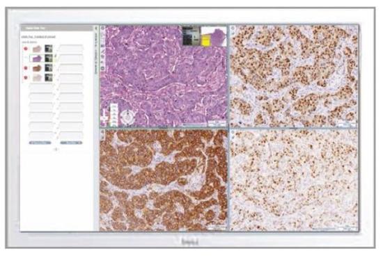
Qualidade acima de tudo o resto
A interface de utilizador intuitiva fornece criação rápida e perfeita de lâminas inteiras, lâminas digitais com alta qualidade de digitalização e resolução com >98 % de taxa de sucesso de digitalização na primeira tentativa.
Tempo curto para ver
O Aperio CS2 digitaliza e movimenta imagens em tempo recorde para análise remota por parte do Patologista. Com um rápido interface de rede de 1GB/segundo, as lâminas digitais estão prontas para análise na sua bandeja de eSlides em 90 segundos.
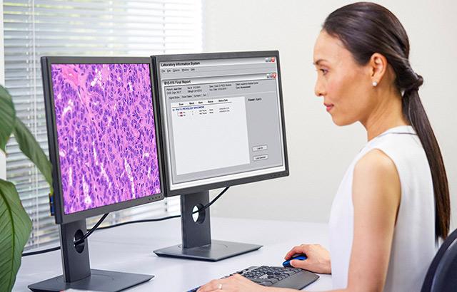
Partilhe em qualquer momento, em qualquer lugar
Aperio CS2 é a ferramenta ideal para preparar lâminas e dados a serem partilhados com outros membros da equipa para colaboração. A avaliação remota de lâminas digitais pode melhorar o tempo de resposta do estudo e a facilidade de acesso para os membros da equipa.
Localizador de tecido
O localizador de tecido Aperio CS2 é muito robusto, mas flexível quando necessário. A área de interesse pode ser ajustada pelo utilizador para otimizar a identificação de tecido em amostras difíceis (como tecido adiposo) ou para evitar detetar tinta ou resíduos de manchas.
"Já não precisamos de criar manualmente experiências de imagem: a poupança de tempo da imagem automatizada é enorme! Além disso, a qualidade consistente das digitalizações, o equilíbrio de brancos e a focagem são impressionantes e facilitam a apresentação de dados a colegas que não estão familiarizados com a histologia."
Fonte: TechValidate. TVID: 1DA-AC0-A5E
PI/Principal, empresa farmacêutica de grande escala
Especificações
GERAL
| Capacidade | 5 lâminas (1 x 3 “), 2 diapositivo (2 x 3") |
| Dimensões | Aproximadamente 12,5 polegadas (L) x 19 polegadas (a) x 20,5 polegadas (D) (318 x 465 x 502 mm) |
| Lentes das objetivas | Olympus UPLXAPO, 20x, 0.80NA, 0.6mm WD, 9mm focal, 26.5FN (digitalização de 40x com comutador de ampliação ótica de 2x) |
| Formato de entrada | Lâminas de microscópio de 1 polegada x 3 polegadas (25 mm x 75 mm) 2 x 3 polegadas (5,08 cm x 7,62 cm) (opcional) |
| Peso | Aproximadamente. 25 kg (55 lbs) |