
Digital Pathology: Take Your Next Step On the Path to Implementation

Digital pathology can help overcome time, productivity, and quality constraints often associated with manual microscopy, by enabling you to work in the digital environment. Additional benefits of implementing digital pathology into your research projects are highlighted below.
Why Go Digital?
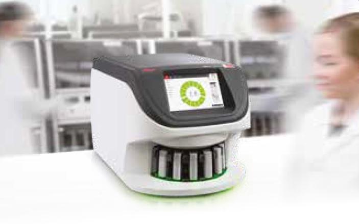
Digital pathology is the practice of converting microscopic glass slides into digital slides that can be viewed, managed, and analyzed on a computer screen or mobile device. Digital pathology is being readily adopted by research organizations to enhance biomarker and drug discovery. Here are the top reasons to implement digital pathology.
Improve Quality & Productivity

Reduced errors:
Helps prevent breaking of precious slides due to excess handling, and need to ship to distant laboratories. Misidentification may also be reduced by barcoding of slides.

More innovation:
Big data provides more research outputs for complex research questions. Allows research groups to collaborate at broader locations. Provides tools for teaching and training at different locations.

Improved analysis:
Improve accuracy in tissue analysis by removing the subjectivity and inter-/ intra-observer variability inherent in manual review, as well as providing a comprehensive, quantitative analysis.

Improved workflow:
Central storage and data management systems for digitized slides enables international collaboration, a more streamlined workflow, and for more flexible work schedules.

A new way to view:
Team annotation of slides. Measure multiple areas of interest with multiple people annotating. Get a dashboard view of all annotations for analysis. Simultaneous view of slides from the same study.
Reduced turnaround time:
Enables immediate access to archive digital slides compared to searching in physical repositories. Also reduces time in retrieving, matching and organizing data within a study.
For research use only. Not for use in diagnostic procedures
About the presenter
Rhian is a Scientist from Swansea University in Medical and Healthcare Studies and was featured in several collaborative publications. Rhian’s research-based background focused on tissue-based pathology in Multiple Sclerosis, primarily using immunohistochemical analysis and in vitro molecular techniques. She spent a short period conducting routine PCR testing for COVID-19 at the end of 2020.
Related Content


El contenido de Leica Biosystems Knowledge Pathway está sujeto a las condiciones de uso del sitio web de Leica Biosystems, disponibles en: Aviso legal.. El contenido, incluidos los webinars o seminarios web, los recursos de formación y los materiales relacionados, está destinado a proporcionar información general sobre temas concretos de interés para los profesionales de la salud y no está destinado a ser, ni debe interpretarse como asesoramiento médico, normativo o jurídico. Los puntos de vista y opiniones expresados en cualquier contenido de terceros reflejan los puntos de vista y opiniones personales de los ponentes/autores y no representan ni reflejan necesariamente los puntos de vista ni opiniones de Leica Biosystems, sus empleados o sus agentes. Cualquier enlace incluido en el contenido que proporcione acceso a recursos o contenido de terceros se proporciona únicamente por comodidad.
Para el uso de cualquier producto, debe consultarse la documentación correspondiente del producto, incluidas las guías de información, los prospectos y los manuales de funcionamiento.
Copyright © 2026 Leica Biosystems division of Leica Microsystems, Inc. and its Leica Biosystems affiliates. All rights reserved. LEICA and the Leica Logo are registered trademarks of Leica Microsystems IR GmbH.