Computational Pathology Solutions from Leica Biosystems

Computational Pathology: Building a Connected Digital Pathology Ecosystem

Turning digital pathology images into actionable insights.

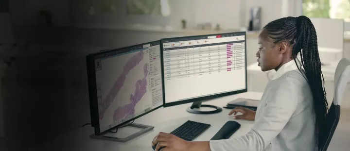
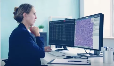
Computational pathology combines whole slide imaging, image management, artificial intelligence, and quantitative image analysis to extract meaningful information from tissue samples. For academic medical centers and research organizations, computational pathology can help improve workflow efficiency, support biomarker discovery, increase reproducibility, and expand access to pathology expertise.

Success depends on more than AI algorithms alone. It requires a connected digital pathology ecosystem that spans the entire workflow, from specimen preparation and slide creation through scanning, quality control, image management, analysis, and collaboration.

Image
halo-ap-Reporting-Options

What Is Computational Pathology?

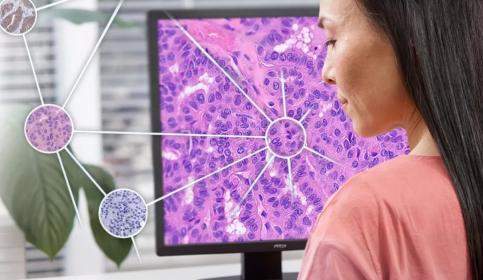
Computational pathology enables pathologists and researchers to extract deeper insights from digital pathology images using AI-powered image analysis and quantitative tissue analytics. These tools help identify patterns, quantify biomarkers, and evaluate tissue characteristics at a scale that would be difficult to achieve through manual review alone.

At the same time, computational pathology depends on the quality of the digital images being analyzed. Consistent specimen preparation, staining, scanning, and quality control are critical for generating reliable data and supporting confidence in downstream analysis. By digitizing routine workflows and enabling algorithm-assisted analysis, laboratories can improve standardization while allowing pathologists to focus their expertise on higher-value research activities.

Image
Digital Pathology
Image
neon motherboard background

The Foundation: Digitization at Scale

As digital pathology adoption grows, laboratories must manage increasing image volumes while maintaining image quality and workflow efficiency. High-throughput whole slide imaging provides the foundation for computational pathology by generating consistent digital datasets that support analysis, collaboration, and AI applications.

Leica Biosystems offers scalable scanning solutions designed to support routine pathology, translational research, and enterprise-wide digital pathology programs.

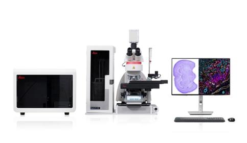
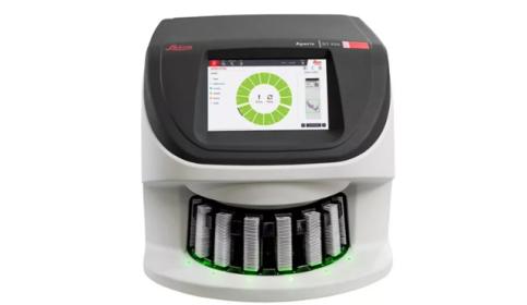
For high-volume laboratories, timely image acquisition is essential to supporting efficient computational pathology workflows. The Aperio GT Elite scanner is designed to deliver rapid, high-quality whole slide imaging at scale, helping laboratories generate the consistent digital datasets required for image management, collaboration, and AI-enabled analysis.

Published performance data indicates scan speeds of up to 103 slides per hour and as fast as 22 seconds per slide, helping laboratories accelerate image acquisition while minimizing manual intervention.

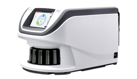
As digital pathology programs expand, laboratories need scalable workflows that can support growing slide volumes while maintaining operational efficiency. The Aperio GT 450 scanner provides a scalable digital pathology platform suitable for routine pathology, translational research, and enterprise deployments.

In a workflow study conducted at NeoGenomics, the Aperio GT 450 scanner demonstrated measurable efficiency improvements compared with the previous-generation Aperio AT2 Scanner, including:

  • 64% improvement in throughput
  • 94% reduction in quality control technician time

Aperio FL Scanning Systems

The Aperio FL slide scanners provide high-quality, high-resolution whole slide images of your research slides. Leverage the flexibility of brightfield, fluorescence, and FISH scanning in a single platform, with 10 or 120 slide capacity options.

For Research Use Only. Not for Use in Diagnostic Procedures.

Learn More

Aperio GT 450 Scanner

Proven technology, with enhanced features, provides efficient workflows and excellent image quality, ensuring seamless integration and secure, optimized delivery of your research.

For Research Use Only. Not for Use in Diagnostic Procedures.

Learn More

Aperio iQC Software

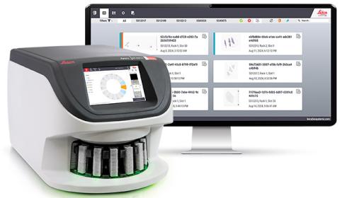
Using AI-powered detection, Aperio iQC Software identifies digital slide artifacts and provides notifications where they're needed—on the scanner console. An intuitive dashboard allows for rapid review and assessment of detected artifacts.

For Research Use Only. Not for Use in Diagnostic Procedures.

Learn More

Aperio GT Elite Scanner

High throughput digital pathology scanner with SmartscanTM technology, delivering speed, intelligence, and certainty in one system.

For Research Use Only. Not for Use in Diagnostic Procedures.

Learn More

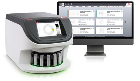
Aperio HALO AP Software

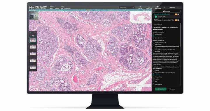
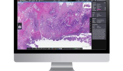
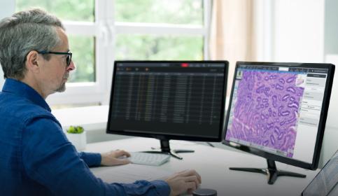
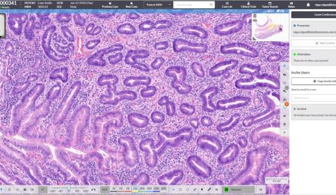
Aperio HALO AP software is designed for efficient on-screen review. It unifies data, whole slide images, and AI-driven analytics into a single, intuitive interface.

Learn More

Aperio AI Store

Aperio AI Store is an analysis platform, seamlessly embedded in Aperio HALO AP software, providing access to a curated menu of leading innovative AI tools. Explore the Aperio AI Store gallery to identify the right solution for your analysis needs.

Learn More

HALO

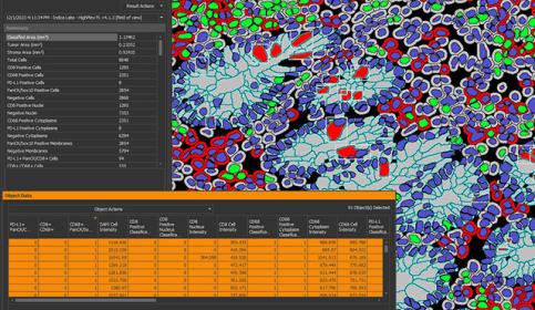
The HALO® platform from Indica Labs provides quantitative and intuitive image analysis for digital pathology. Choose from a broad range of brightfield, fluorescent and multiplex image analysis tools to complement your research.

Learn More

Image
Aperio HALO AP DX brochure

Reliable Computational Pathology Requires Quality Control

Artificial intelligence is only as reliable as the data used to train and deploy it. Digital pathology artifacts introduced during specimen preparation, staining, coverslipping, scanning, or image acquisition can affect image quality and downstream analysis.

To help laboratories identify image quality issues earlier in the workflow, Leica Biosystems offers Aperio iQC software, an AI-powered quality control solution. Integrated directly with the Aperio GT scanner family, Aperio iQC software automatically detects six common image issues during scanning and alerts users while slides remain on the scanner, helping support more consistent digital pathology outputs. 

A study conducted at Heidelberg University underscored the value of automated quality control in digital pathology workflows. Published performance results demonstrated:

  • Reduced reviewer-specific time, including a 68.6% reduction for digital pathology technicians (17.3 minutes per case) and a 34.2% reduction for pathologists (3.4 minutes per case).
  • Detection of up to 24% more artifacts with Aperio iQC software compared with manual review by histotechnicians, supporting consistent, reproducible, standardized results.
  • Reduced quality control costs by automating repetitive review tasks, with manual WSI quality control estimated at $1 per slide in technician time alone.

These findings highlight how AI-assisted quality control can improve efficiency and reduce the time required for individual slide review.
 

From Digital Images to Quantitative Insight

Image management

Once slides have been digitized and quality checked, institutions need efficient ways to store, access, review, and share digital images. Centralized image management helps drive collaboration, remote review, education, and enterprise-wide access to pathology data.

The Aperio HALO AP image management system pairs with the Aperio GT Elite, Aperio GT 180, and Aperio GT 450 scanners to create a connected workflow that unifies digital slides, case information, annotations, and collaborative review. By bringing these elements together in a single environment, Aperio HALO AP software helps support efficient image management across digital pathology programs.

Quantitative analysis

Computational pathology extends beyond image viewing. Quantitative image analysis tools enable researchers to generate objective measurements and transform whole slide images into datasets that support biomarker discovery, translational research, spatial biology, and pharmaceutical development.

Image
Aperio-HALO-AP-Case-Conferencing-presenter
Image
cpath-ai-ecosystem-1100x600

Accessing an Expanding AI Ecosystem

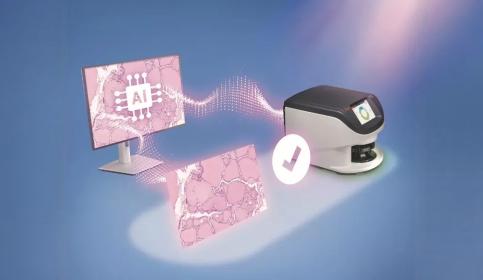
As computational pathology matures, laboratories need flexible ways to evaluate and adopt specialized AI applications without disrupting established workflows or managing separate software environments. A connected ecosystem enables organizations to expand capabilities while maintaining consistency across imaging, review, and analysis activities.

The Aperio AI Store helps support this approach by providing Aperio HALO AP software users with access to a curated ecosystem of digital pathology AI applications from multiple providers. Available within a unified interface, these tools support a variety of tissue analysis, biomarker assessment, and computational pathology use cases.

Understanding the ROI of Computational Pathology

The value of computational pathology extends beyond faster scanning or image storage. By connecting digitization, quality control, image management, and analysis within a unified workflow, organizations can realize value across operational, clinical, and research activities. Potential areas of return on investment include:

  • Reduced manual slide handling and transportation
  • More efficient quality control workflows
  • Increased scanner throughput
  • Improved access to subspecialty expertise
  • Faster consultation workflows
  • Standardized biomarker quantification
  • Scalable support for research programs
  • Reduced variability in image review processes
  • Improved utilization of pathology resources

By automating routine tasks, computational pathology helps laboratories make better use of pathology expertise, enabling pathologists to focus on interpretation, collaboration, education, and research.

Image
Computational Pathology ROI Standard

Key Takeaways

  1. Computational pathology depends on high-quality digital images, structured workflows, and scalable analysis capabilities.
  2. Whole slide imaging platforms such as the Aperio GT Elite scanner, Aperio GT 450 scanner, Aperio GT 180 scanner, and Aperio FL platform provide the digital foundation required for computational pathology.
  3. AI-powered quality control tools such as Aperio iQC software can help reduce manual review burden while improving image consistency.
  4. Aperio HALO AP software, HALO image analysis platform, and the Aperio AI Store help transform images into quantitative insights and deploy AI tools within routine workflows.
  5. The most successful computational pathology programs integrate scanning, quality control, image management, analysis, and collaboration into a connected ecosystem.
     

Have Questions?

Computational pathology requires more than a scanner or an AI algorithm. It requires a connected digital ecosystem capable of supporting image acquisition, quality control, image management, quantitative analysis, and future AI innovation.

Whether you are building a new digital pathology program, expanding research capabilities, or evaluating AI deployment strategies, Leica Biosystems can help you assess workflow requirements and integration opportunities.

Let us help you start your computational pathology journey.

 Click below to request a live demonstration, schedule a consultation, or request pricing. 

Talk to an Expert Today

Resource Hub

Explore more ways to learn about computational pathology

Use these resources to learn how scanning technology, automation, workflow design, and software can work together to enable computational pathology.

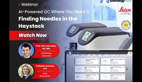
AI-Powered QC Where You Need It: Finding Needles in the Haystack

Learn how AI-powered QC helps labs identify the minority of slides with artifacts, freeing up technician time to focus on the critical few.

Aperio GT 450 Improves Throughput by 64% and Reduces QC Tech Time by 94% in Workflow Study

This white paper evaluates ways to scale up digital pathology operations to keep up with increasing demand, assessing the performance of the Aperio GT 450 versus Aperio AT2.

Aperio iQC Software – AI-Powered Whole Slide Imaging Quality Control

Using AI-powered detection, Aperio iQC Software identifies digital slide artifacts and provides notifications where they're needed - on the scanner console. An intuitive dashboard allows for rapid review and assessment of detected artifacts.

Glossary

Computational Pathology
The application of digital imaging, AI, and quantitative analysis to pathology data.

Whole Slide Imaging (WSI)
The process of scanning an entire glass slide to create a high-resolution digital image.

Digital Pathology
The practice of managing, viewing, interpreting, and sharing pathology specimens digitally.

AI-Powered Quality Control
Automated identification of image artifacts and quality issues using machine learning algorithms.

Biomarker Quantification
Measurement of cellular or tissue characteristics to support research or clinical assessment.

Image Management System (IMS)
Software used to organize, store, retrieve, and collaborate around digital pathology images.

Spatial Biology
The study of the organization and interaction of cells within their tissue environment.

Frequently Asked Questions

What is the difference between digital pathology and computational pathology?

Digital pathology focuses on digitizing and viewing pathology slides. Computational pathology builds upon digital pathology by applying AI, machine learning, and quantitative image analysis to generate additional insights from digital images.

Why is image quality important for computational pathology?

Poor image quality can affect algorithm performance, quantitative measurements, and workflow reliability. Computational pathology requires consistent, high-quality images to produce reproducible results.

How does AI improve pathology workflows?

AI can assist with quality control, image review, biomarker quantification, tissue classification, workflow prioritization, and quantitative analysis. These capabilities help reduce manual effort while increasing consistency.

What infrastructure is required to implement computational pathology?

Successful programs typically require slide scanning, image storage, image management software, quality control processes, network infrastructure, user training, and analysis platforms.

Can computational pathology support research applications?

Yes. Computational pathology is widely used in translational research, biomarker discovery, immuno-oncology, spatial biology, pharmaceutical development, and multicenter collaboration programs.

How can laboratories evaluate the ROI of computational pathology?

Organizations often evaluate throughput improvements, labor efficiency, quality metrics, consultation turnaround times, access to expertise, scalability, and support for research productivity.

How does Leica Biosystems support computational pathology adoption?

Leica Biosystems provides an integrated ecosystem spanning slide scanning, AI-powered quality control, image management, quantitative image analysis, and access to third-party AI applications within connected workflows.