
 -
-
 Logout



Mindpeak PanTumor H&E
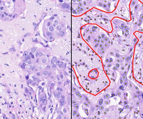
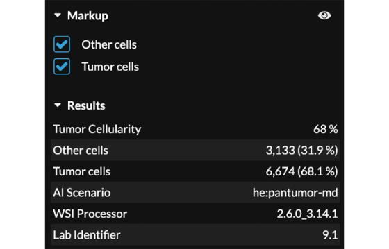
Mindpeak PanTumor H&E is an AI-powered application for automated tissue segmentation of H&E-stained whole slide images across tumor types. Built on a pan-cancer foundation model, it segments the whole slide into tumor, stroma, necrosis, and normal tissue when the whole slide image is opened in a research workflow. The application counts the tumor and other cells and outputs tumor cellularity as a percentage—no region drawing and no manual counting. The proposed regions can then be reviewed and adjusted.
For Research Use Only. Not for use in diagnostic procedures.


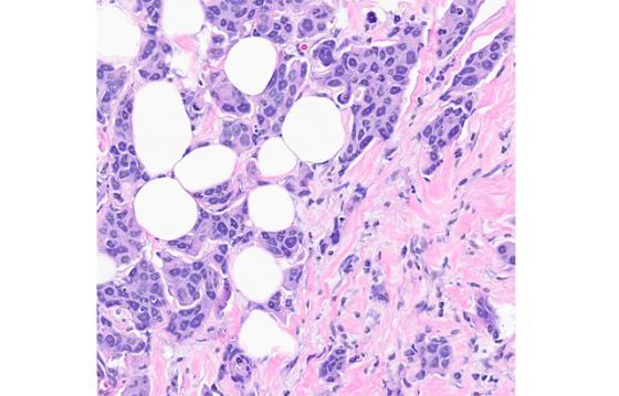

 Zoom image
 0-Click Pan-Tumor Segmentation
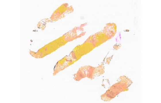
Almost every downstream task in pathology starts with the same question: where is the tumor and how much of it is there? When a compatible H&E whole slide image is opened, a single pan-cancer foundation model answers it automatically across the entire slide—no region drawing, no manual outlining. The tissue is segmented into tumor, stroma, necrosis, and normal tissue and rendered as a color-coded overlay covering every tissue fragment on the section, including small or fragmented tissue pieces that may be overlooked during manual review. The result is a map of the whole slide rather than an impression derived from the portion examined most closely. Because one model produces all four classes in a single pass, the map is generated consistently. Because cellularity is counted rather than estimated, the scoring value is based on the underlying tissue.
You Keep Control of the Boundary
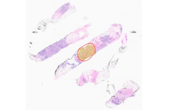
Segmentation is a proposal, not a verdict. Using the standard annotation tools, the regions the model proposes can be accepted, rejected, edited, or added to, and the cell counts and cellularity follow the regions as they are adjusted. That keeps the user in control of the boundary that matters most, which is the boundary between tumor and non-tumor tissue. Because the adjustment happens on regions rather than on the underlying image analysis, a corrected map is available immediately, and the rationale behind the scoring remains visible. Every adjustment is made using tools that users already know from routine annotation work, so nothing new has to be learned to correct a boundary. The user reviews the result and remains responsible for the final analysis.
A Foundation for Downstream Workflows
A tumor map is rarely the final output. Rather, segmentation provides the foundation for downstream research workflows that would otherwise require manual annotation. These workflows include guiding microdissection and tumor enrichment prior to molecular analyses, limiting IHC quantification to tumor regions to prevent signal dilution from adjacent tissue, and independently quantifying tumor, stromal, and necrotic content. One automated analysis of the H&E therefore replaces multiple manual outlining steps, and every downstream workflow starts from the same delineation rather than from a fresh assessment. For molecular analysis in particular, tumor cellularity is a key parameter for the assay and knowing it before a sample is submitted can reduce the likelihood of tissue containing insufficient tumor content.
Consistent Delineation
Outlining tumor by hand is slow and subject to variability: two resaerchers draw different boundaries on the same section, and the same reader may draw a different boundary on a different day. Anything computed from that boundary inherits that variation, which can be important when a tumor content measurement is used to determine whether a sample proceeds to molecular analysis. Mindpeak PanTumor H&E applies the same criterion consistently across the whole slide, so the delineation does not depend on who opened the whole slide image or how long they spent on it. The benefit is most apparent in challenging sections: fragmented biopsies and tumors with diffuse borders are exactly the sample where two researcher are least likely to draw the same line. Where the model and the user disagree, the user adjusts the regions and the figures follow.
Counts and Cellularity, Fully Traceable
Counts and cellularity, traceable to the map behind them. Analyses are returned as the number of tumor cells, the number of other cells, and tumor cellularity expressed as a percentage of all counted cells. Because the counts are displayed next to the percentage derived from them, a reviewer can verify any reported value against the cells that produced it. Individual tissue classes can be shown or hidden during review, so the tumor delineation can be inspected on its own before the figures are accepted. The panel also records the provenance of each analysis: the AI scenario applied, the processor version, and the laboratory identifier. The application supports whole slide images from a range of scanners.
An H&E-stained tumor whole slide image is opened, and segmentation starts automatically, with no region drawn and no tissue outlined by hand. Within seconds, the whole slide is divided into tumor, stroma, necrosis, and normal tissue in the background, and the tumor and other cell counts appear alongside the tumor cellularity derived from them. The video then shows a proposed region being adjusted with the standard annotation tools and the figures following the change. Tissue classes are shown and hidden so the tumor delineation can be inspected on its own. At no point in the workflow is a region drawn before the segmentation begins.
Specifications
| Tissue type | Multiple solid tumor types; formalin-fixed, paraffin-embedded sections stained with hematoxylin and eosin |
| Stain: | Hematoxylin and eosin (H&E) |
| Tissue classes returned: | Tumor; stroma; necrosis; normal tissue |
| Required image resolution | 0.5 µm/pixel plus/minus 10% (20x equivalent) |
| Outputs | Tumor cell count; other cell count; tumor cellularity as a percentage of all counted cells; tissue-class segmentation overlays |
| Analysis mode | 0-click whole slide segmentation, with proposed regions reviewable and adjustable using the standard annotation tools |