








Lunit-SCOPE-PD-L1-CAL10-NSCLC-Positive-Negative
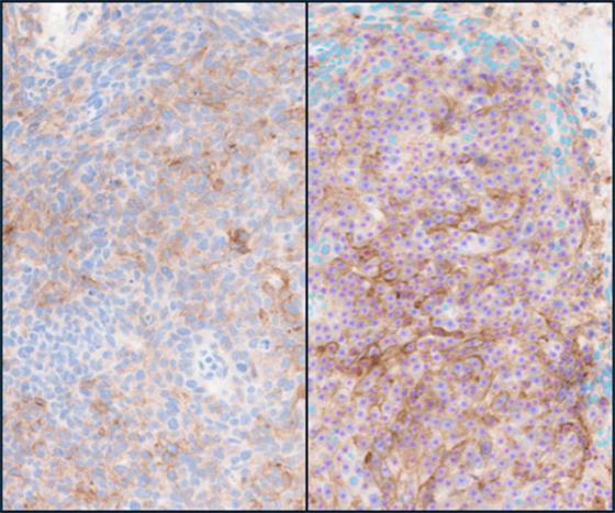
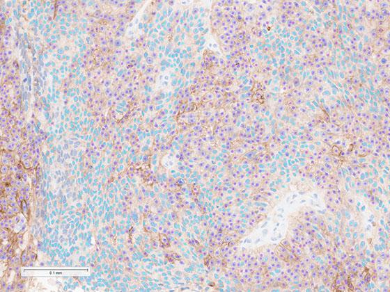
PD-L1 (CAL10) IHC digital slide (left) with Lung PD-L1 AI markup (right) identifying negative tumor cells (cyan), and positive tumor cells (magenta).

Lunit SCOPE PD-L1 CAL10 NSCLC

Lunit SCOPE is an artificial intelligence (AI)-based digital pathology image analysis software that empowers researchers to improve accuracy, scoring agreement, and workflow efficiency. Lunit SCOPE PD-L1 CAL10 NSCLC assists in the detection and quantification of PD-L1 expression in non-small cell lung cancer (NSCLC) tissue stained with Leica Biosystems’ PD-L1 (CAL10) primary antibody.

Lunit SCOPE PD-L1 CAL10 NSCLC automates tumor proportion score (TPS) calculations and marks up images with tumor detection and cell phenotyping based on PD-L1 (CAL10) staining. The AI-powered algorithm seamlessly integrates with Leica Biosystems’ staining and digital pathology ecosystem to streamline workflows.

Activate AI Now

Contact us

Lunit SCOPE PD-L1 CAL10 NSCLC is For Research Use Only. Not for use in diagnostic procedures.

Lunit SCOPE PD-L1 CAL10 NSCLC is accessed via the Aperio HALO AP image management system.

Lunit-Black-logo

Consistent Scoring for PD-L1 Research

  • AI/ML-Based Scoring: The application utilizes robust artificial intelligence and machine learning algorithms, trained on a broad spectrum of PD-L1 (CAL10) stained tissue samples. This ensures accurate TPS quantification, particularly around critical cutoffs (1% and 50%), addressing common interpretation challenges such as borderline cases, macrophage staining, and human subjectivity.
  • Automated TPS Scoring: By automating TPS scoring, the application saves valuable reading time, especially for complex cases. This efficiency allows researchers to focus on higher-value tasks and enhances productivity.
  • Seamless Digital Pathology Integration: Designed for the Leica Biosystems digital pathology ecosystem across digital slide scanner, image management, and AI image analysis. Researchers benefit from streamlined workflow integration that enables reproducibility across multiple study sites.
Image
Lunit-SCOPE-PD-L1-CAL10-NSCLC-PD-L1-Scoring-Range
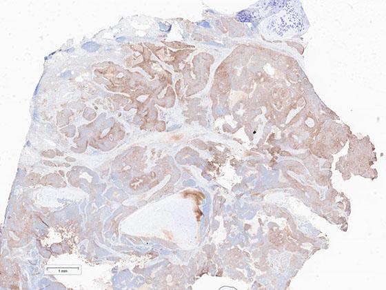
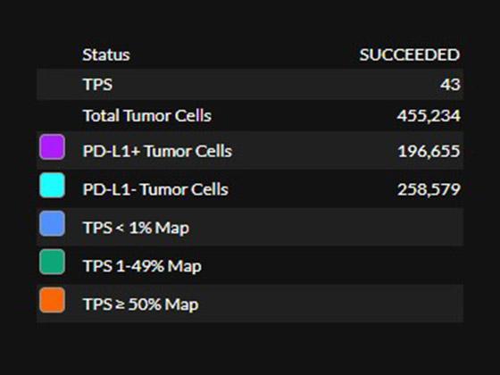
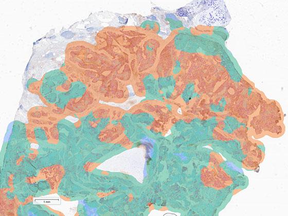
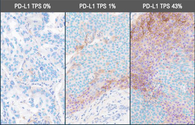
PD-L1 (CAL10) IHC digital slides with PD-L1 AI markup at 0% TPS (left), 1% TPS (middle), and 43% TPS (right). Negative tumor cells (cyan), positive tumor cells (magenta).

Lunit SCOPE PD-L1 CAL10 NSCLC Demonstration in Aperio HALO AP Image Management System

Explore the future of pathology with Lunit SCOPE PD-L1 CAL10 NSCLC. The PD-L1 image analysis algorithm is designed for intuitive use within the Aperio digital pathology environment. The algorithm automatically identifies regions of interest and applies AI-powered image analysis to calculate the tumor proportion score (TPS), presenting analysis in a clear, interactive dashboard.

Specifications

Scanner: Compatible file types include Leica Biosystems (.svs), Philips (.iSyntax, .tiff), 3D Histech (.mrxs), Hamamatsu (.ndpi) and other scanners at x40 magnification
IMS: Leica Biosystems’ Aperio HALO AP
Algorithm: Lunit SCOPE PD-L1 CAL10 NSCLC
Assay: Leica Biosystems PD-L1 CAL10
Tissue Type: NSCLC
Scoring: TPS
Regulatory status: Research Use Only (RUO)