
 -
-
 Logout



DiaKwant™ HER2 Breast
Seven membrane categories, read cell by cell. Zero click, zero annotation, zero manual counting: when you open the slide in Aperio HALO AP Software, DiaKwant™ AI has already framed the invasive breast carcinoma, read each tumor cell membrane and computed the HER2 score (0, 1+, 2+, 3+), with the proportion of cells in each category. A heatmap shows how membrane staining varies across the tumor, so the score stays traceable to the tissue. You stay in control. The AI handles the count.
For Research Use Only. Not for Use in Diagnostic Procedures.

 Zoom image
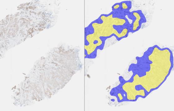
 Region detection and counting are performed automatically
DiaKwant™ AI selects the invasive tumor on the entire slide with no region to draw. In situ components and control areas are excluded from analysis.
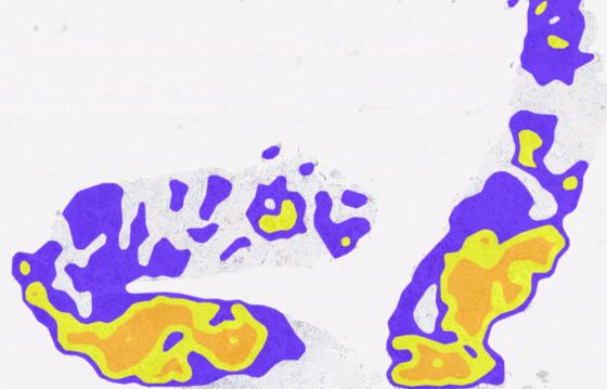
HER2 membrane category markup across the invasive tumor (blue: no membrane staining, yellow: weak staining).
Identify the cells of interest
Each tumor cell membrane is read for completeness and intensity. The AI handles the count you would do manually, on the entire slide.
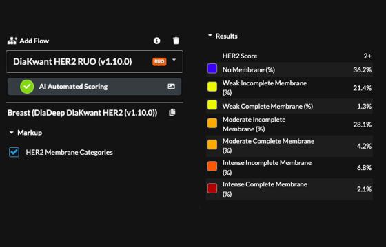
Quantify HER2: a HER2 score (0, 1+, 2+, 3+) with the proportion of tumor cells in each of seven membrane categories:
No Membrane
Weak Incomplete Membrane
Weak Complete Membrane
Moderate Incomplete Membrane
Moderate Complete Membrane
Intense Incomplete Membrane
Intense Complete Membrane
DiaKwant™ HER2 results panel: HER2 score and the percentage of tumor cells in each of the seven membrane categories.
See how staining varies
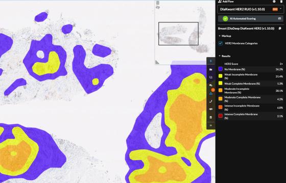
A heatmap of membrane intensity and completeness across the invasive tumor. Switch it on and off to see the tissue underneath.
DiaKwant™ HER2 in Aperio HALO AP Software: membrane category heatmap with the results panel.
DiaKwant HER2 Demonstration in Aperio HALO AP Image Management System
DiaKwant HER2 is a DiaDeep algorithm integrated within the Aperio HALO AP digital pathology platform to deliver accurate HER2 quantification. It automatically analyzes the whole slide to evaluate membrane staining completeness and intensity, with no manual annotation or selection of invasive tumor regions required from the pathologist.
After selecting a HER2 slide, the pathologist can review the tissue in detail. A heatmap overlay highlights membrane staining variations across the invasive tumor, from no staining to intense complete membrane staining. The heatmap can be switched on or off at any time, allowing the pathologist to compare the algorithm's output with the underlying tissue morphology.
At the end of the analysis, the pathologist obtains:
- The HER2 score, an IHC category (0/1+/2+/3+).
- The proportion of tumor cells in each membrane staining category, supporting understanding of the score.
- A heatmap of membrane staining intensity and completeness across the tumor.
DiaKwant also offers automated quantification for PD-L1, Ki-67, ER, PR, and mitosis.
Specifications
| Algorithm | DiaKwant HER2 Breast |
| Supported image formats | SVS, DICOM |
| Deployment | cloud (Software as a Service) or on-premise |
| Outputs | HER2 score (0, 1+, 2+, 3+), proportion of tumor cells per membrane category, heatmap |
| Regulatory status | Research Use Only (RUO) |