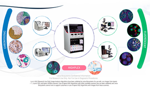
IHC & Multiplexing
Researchers need clear results to discover new treatments. BOND RX fully automated research stainers provide the flexibility you need to explore new possibilities, accurate results to ensure nothing is missed, and rapid, cost-effective operation, so you can perform more tests.
Automated, Multiplexed Co-detection of RNA and Protein
Julia Jones, Scientific manager, Histopathology/ISH Core Facility, Cancer Research UK Cambridge Institute, University of Cambridge
Learn more from subject-matter experts from the Cancer Research UK Cambridge Institute, who will share results using chromogenic methods such as integrated co-detection workflow for RNAscope in situ hybridization assays.

Flex Your Plex
Multiplexing enables a paradigm shift for ISH and immunohistochemistry (IHC). It provides a more complete picture of what is happening at a cellular level, showing spatial arrangements and protein/DNA/RNA interactions all on the same slide and sample.
Enhanced Automated Multiplex Capabilities on Leica Biosystems BOND Research System
Damian Cockfield Global Product Manager - BOND, Leica Biosystems
Dean Talia PhD, Senior Scientist, Leica Biosystems
There is an ever-increasing demand to derive more information from limited research samples. Establishing expression profiles of the tumor microenvironment can be achieved via multiplex staining now with chromogenic detection. Discover how the new features of the Leica BOND research system and new assays combine to support automated 6-plex chromogenic multiplexing.
Multiplex User Meeting – Virtual Event Success
Rhian Evans, Ph.D., Scientist
The first virtual event of the year recaptured the conference atmosphere at the Multiplex User Meeting. With Leica Biosystems total multiplex solutions and trusted partners, advances in multiplex immunohistochemistry (IHC) were innovatively showcased to customers via live presentations, on-demand webinars, virtual exhibition hall, networking room and, virtual lab tour!
Selecting the right detection for your immunohistochemistry (IHC)/ISH project (Fluorescence vs Chromogenic Staining)
Shenna Washington, Sr. Field Applications Specialist, Leica Biosystems
Choosing the appropriate detection method for tissue targets will allow researchers to produce the most compelling visual images to support their research goals.

Dissection of the Melanoma Microenvironment via High-throughput Multiple Iterative Labeling by Antibody Neodeposition (MILAN)
Prof Dr. Francesca Bosisio Assistant Professor, University of Leuven (KU Leuven), Department of Imaging & Pathology, Translational Cell & Tissue Research
Asier Antoranz Post Doc Bioinformatician, University of Leuven (KU Leuven), Department of Imaging & Pathology, Translational Cell & Tissue Research
In this webinar, Prof. Francesca Bosisio and Asier Antoranz, Assistant Professor and Postdoctoral Bioinformatician at University of Leuven, provide a practical example of what digital pathology can achieve, illustrating how high-dimensional single-cell multiplex analysis using multiple iterative labelling by antibody neodeposition (the MILAN technique) can characterize the immune landscape in primary melanoma.

Tips & Tricks to Multiplexing: How to Choose Chromogen Colors for Multiplex and Detection Systems for Multiplex Assays
Rhian Evans Ph.D., Scientist
Learn more about multiplexing and explore these educational videos and infographics to understand how to choose chromogen colors for multiplex, as well as the different detection systems available for multiplex assays.
Tips & Tricks to Multiplexing: Top 5 Reasons to Multiplex and Chromogenic versus Immunofluorescent Detection
Rhian Evans Ph.D., Scientist
Multiplexing is an important tool for simultaneous detection of multiple markers within a single tissue section. These educational infographics highlight the five key reasons to multiplex, as well as the main considerations for choosing chromogenic or immunofluorescent detection.
Spatial Biology
Shubham Dayal, Senior Medical Writer, Medical and Scientific Affairs, Leica Biosystems
Jack Heath, Medical Science Liaison, Ph.D.
This article is the third piece of a four-part series including articles titled “What are Life Sciences,” “Translational Research,” “Spatial Biology/Research” (current feature), and “Biomarkers in Translational/Spatial Research.”
For Research Use Only. Not for use in Diagnostic Procedures.
Featured Life Sciences Products
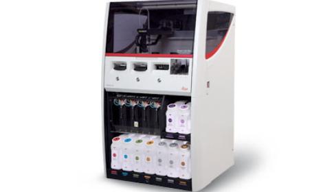
BOND RX
The BOND RX high-throughput research stainer delivers speed and flexibility for IHC, ISH, and multiplexing staining with either chromogenic or fluorescence markers.
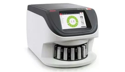
Aperio GT 450 Scanner
Proven technology, with enhanced features, provides efficient workflows and excellent image quality, ensuring seamless integration and secure, optimized delivery of your research.
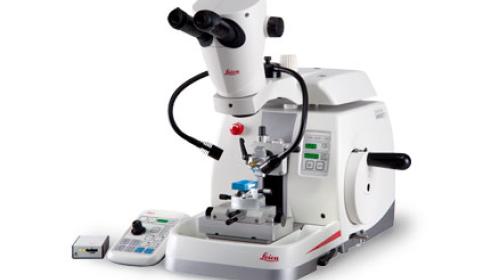
HistoCore NANOCUT R
When your microtomy involves diverse samples in order to discover new breakthroughs in your research projects, then NANOCUT R is the optimal solution for you.
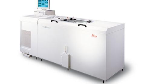
Leica CM3600 XP
Fully computerized cryomacrotome for whole‐body sectioning of small animals.