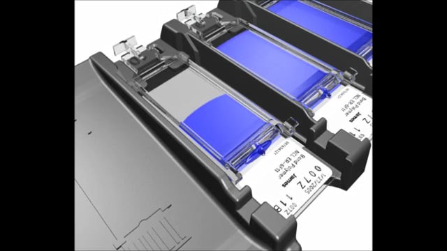

概览

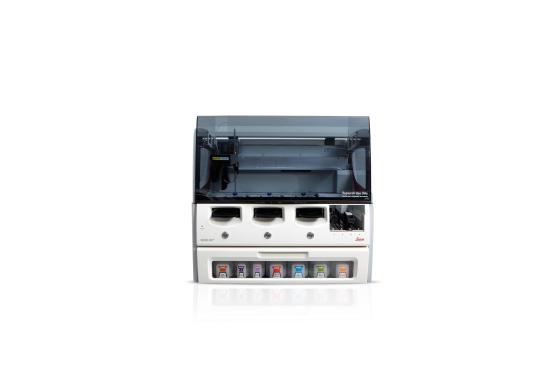
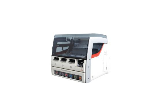
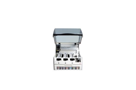
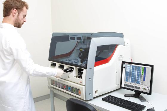
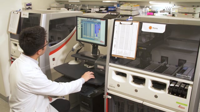
帮助学术研究人员取得更出色的成就:BOND RXm – 全自动高级染色

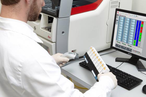
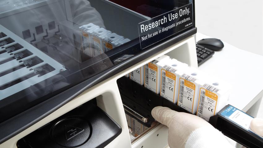
BOND RXm 可实现 IHC、ISH 和新兴检测的完全自动化,让您享受更多的学术研究时间。无与伦比的程序灵活性和完全开放式试剂,意味着您可以全面控制染色过程。三个独立的玻片架还可确保随心按照您自己的时间表运行玻片染色。

联系我们 询价。 请致电 +65 6537 6815 与销售人员取得联系。

BOND RXm 仅可用于科研用途,不适用于诊断用途。


特性

温度工具箱
时间
灵活性
一致性

BOND RX 软件 7.0 版本

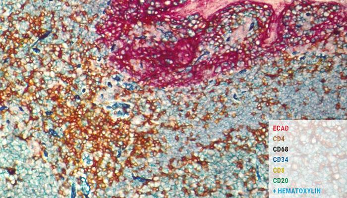
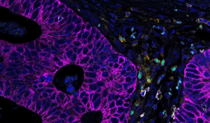
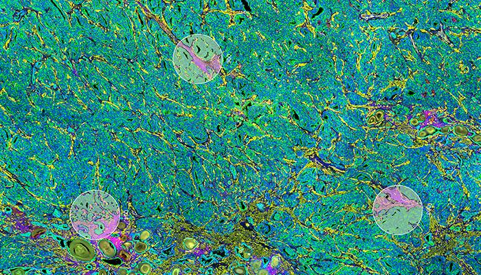
BOND RX 7.0 软件支持改进的显色和荧光多重染色功能,可在单张玻片上显示多达 6 个不同的标记。

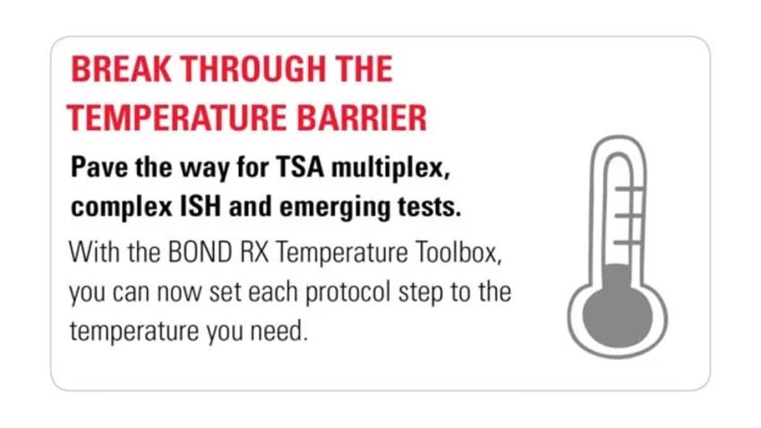
突破染色屏障

每张玻片上的标记越多,您就能更快到达突破点,同时减少使用的珍贵组织。您可以完全定制化程序,包含从脱蜡到复染之间的所有步骤,从每个标本获得更多的数据。

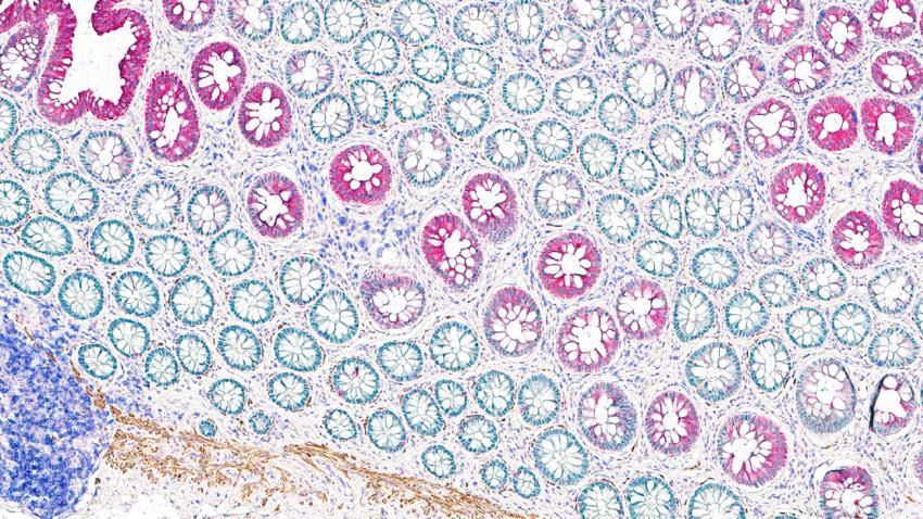
  • 通过多重染色解锁细胞秘密,在单张玻片上实现多个目标的可视化
  • 通过设计您自己的定制化检测系统和理想程序,挖掘下一个突破性试验方法
Image
Break_Through_the_Staining_Barrier

解锁细胞秘密

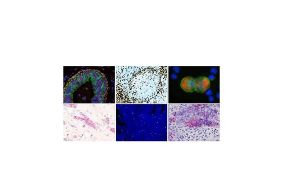
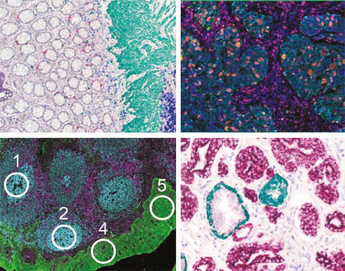
使用科研级 BOND 染色机进行多重染色,从而

  • 发现空间关系
  • 确定细胞表型
  • 借助单个组织标本获取更多的数据

由广泛的技术合作伙伴提供支持


Image
Unlock_Cellular_Secrets

设计未来的突破性检测

自由探索

  • 最多可染色 6 个标记
  • DNA、RNA 和蛋白质
  • FFPE 和 CTC
  • 显色、荧光和测序
Image
Design the Next Breakthrough Assay

探索颜色选项

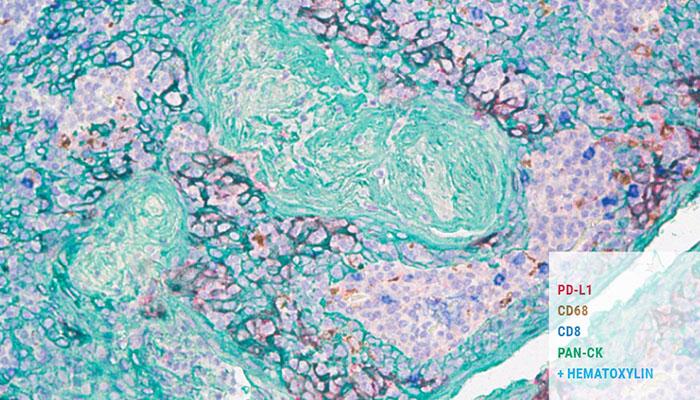
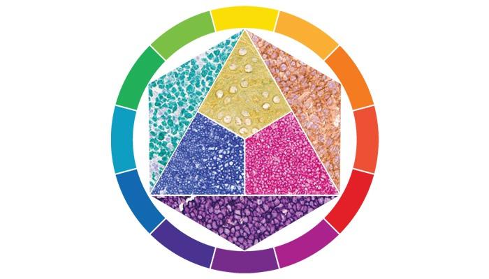
每张玻片最多可选择 6 种显色剂;可即插即用或自行选择。

让仪器混合专为 BOND 染色机设计的显色剂。使用我们的即插即用显色剂,只需扫描并装载至 BOND 仪器中即可。
Image
Explore the Color Options

为您的目标量身定制

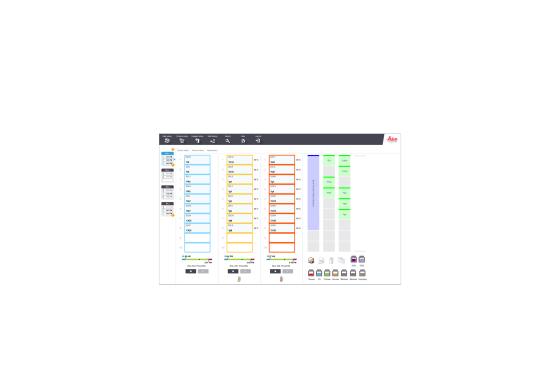
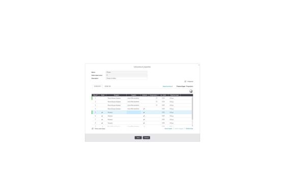
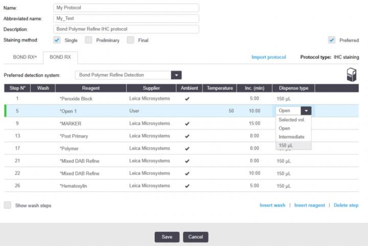
BOND RX 软件 7.0 版本可为设计理想的程序带来灵活性

  • 每张玻片最多可选择 6 个标记
  • 创建您自己的检测系统
  • 完全定制您的所有研究程序
  • 更改时间、温度和分配类型
Image
Tailor to Your Target

获得最新研究进展

Leica Biosystems 与众多开放式创新 (OI) 合作伙伴合作。因此,才能为您的研究提供许多灵活的解决方案。对于我们与 OI 合作伙伴联合开发的所有程序和程序模板,均可于 BOND RX 和 RXm 上获取,以便您在最短的时间内获得最新研究进展。

完全集成式 RNA ISH 解决方案
合作伙伴 - ACD

借助高通量的染色技术,可在每次运行中最多完成30张玻片操作,如果是单面,则每次运行需要11个小时,如果是双面,则每次运行需要14个小时。此解决方案具有以下特性:

  • 高特异性 – 此解决方案使用双 z 探针策略和同步背景抑制策略放大信号,以确保与目标发生特异性结合。
  • 灵敏且可定量 – 以单细胞分辨率实现单 RNA 分子检测。
  • 形态 – 此解决方案可在信息完整的组织结构中提供细胞特异性表达信息。
  • 通用性 – 几乎可用于任何基因、物种或组织。

观看合作视频 - ACD RNAscope 了解更多

Image
A_Fully_Integrated_RNA_ISH_Solution_Partner_ACD

FFPE 的蛋白质和 RNA 的高复杂空间结构分析
合作伙伴– NanoString

将标准的免疫荧光技术与数字光学条形码技术相结合,GeoMx™ 数字空间分析仪可实现蛋白质和 RNA 高度复杂的空间结构分析。GeoMx 检测可从单个 FFPE 或新鲜冷冻的组织切片进行成像和分析。

  • 在空间范围内量化多达 96 种蛋白质和 1000 多种 RNA 靶标
  • 以组织中相关稀有细胞或隔室特定区域为重点
  • 通过非破坏性处理保留珍贵样品

了解更多

Image
High Plex, Spatial Profiling of Proteins and RNA from FFPE Partner – NanoString

将 CTC 分析应用于实验性病理实验室
合作伙伴 - Rarecyte

AccuCyte-Cytefinder 系统与 BOND RX 结合,可在显微镜下显示循环肿瘤细胞。该系统可在下游基因组分析中,对靶细胞进行单细胞修复。
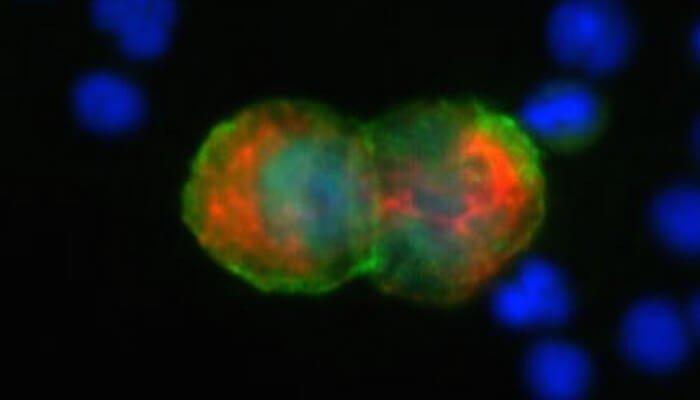
  • AccuCyte 从全血中收集有核细胞,包括循环肿瘤细胞。
  • CyteSealer 在血块黄层与红细胞间创建一道屏障,将收集的细胞密封起来。
  • CyteSpreader 确保每次将精确数量的有核细胞分散到各玻片上。
Image
CTC_Analysis_For_Experimental_Pathology_Labs_Partner_Rarecyte

全自动操作,实现单步多重分析
合作伙伴 - Ultivue

在 6 小时内,使用 BOND RX 最多可制作 30 块五色玻片,在实现单步多重分析的同时可保持组织形态和完整性。因此,研究员既可节省完成高难度手工技术的时间,又可腾出更多时间重点研究下一个突破点。
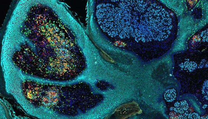
  • 通过高效能组织生物标记物多重检测,在肿瘤微环境内使用空间相关性调查分析细胞表型
  • 采用 DNA 条形码抗体和唯一互补的萤光 DNA 条形码,通过单一的混合抗体和孵育步骤对单块玻片进行多重分析
  • 保持标本的完整性,从而在不损害组织或不相互干扰的情况下做进一步分析
  • 使用 Aperio VERSA 和 Aperio 细胞 IF 算法捕获图像和分析
Image
Single_Step_Multiplexing_Fully_Automated_Partner_Ultivue

Fully automated Navinci in situ proximity ligation assays: Naveni® kits for BOND RX/RXm research stainer
Partner - Navinci

Navinci in situ proximity ligation assays offer novel technology with a choice of chromogenic and fluorescent readout on the BOND RX and BOND RXm automated research staining platform by Leica Biosystems.
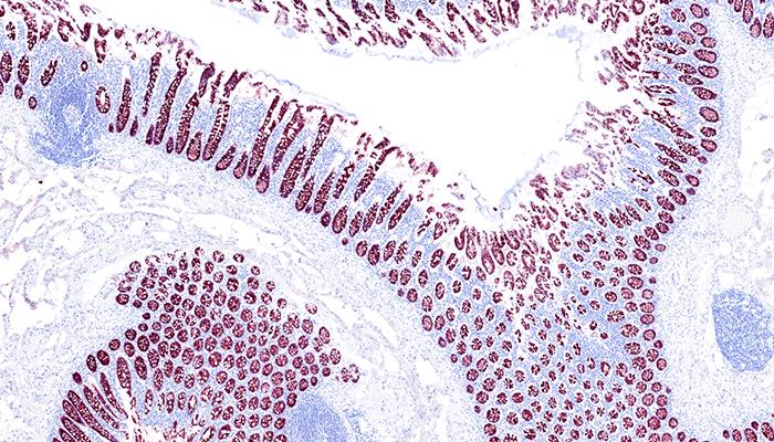
  • Detect protein-protein interactions and post-translational modifications efficiently using the BOND RX research stainer, ensuring high precision and accuracy
  • Visualize specific protein targets like PD1/PD-L1 while preserving intact cell and tissue morphology, providing a holistic understanding of spatial protein relationships
  • Ready-to-use kits optimized for FFPE samples and reagents sufficient for 30 FFPE tissue slides

Learn More

Image
Navinci-Our-Innovation-Partners-HUB-700pxX400px

SignalStar® Multiplex IHC Fully Automated on the BOND RX and BOND RXm Research Staining Platforms
Partner - Cell Signaling Technology (CST)

Streamline multiplex spatial image generation and analysis with flexible, rigorously validated antibody panels and optimized protocols.
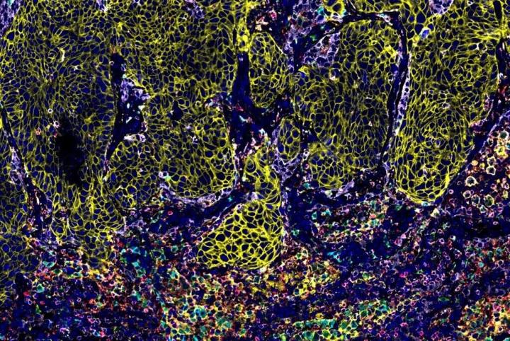
  • Stain 10 8-plex samples in two days with validated protocols on the BOND RX research stainer
  • Remove up to 12 hours of hands-on time from the SignalStar workflow with automation
  • Build panels in four simple steps with an expanding menu of oligo-conjugated, IHC-validated primary antibodies

Learn More

Image
Cover-image-1_1

Fully Automated HCR™ Assays on the BOND RX and BOND RXm Research Staining Platforms
Partner - Molecular Instruments (MI)

MI's HCR™ assays deliver high specificity and resolution for RNA and protein detection in FFPE tissue samples on the BOND RX and BOND RXm automated research staining platforms.
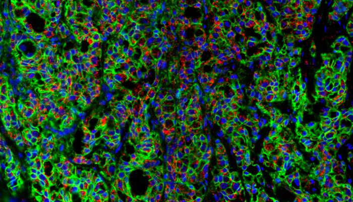
  • Streamline automated RNA and protein workflows on the BOND RX and BOND RXm research staining instruments
  • Stain up to 30 slides in under 10 hours on the BOND RX and under 11 hours on the BOND RXm research staining platform using pre-loaded protocols
  • HCR™ Pro RNA-ISH enables entirely protease-free detection of diverse RNA targets, including microRNAs, splice junctions, lncRNAs, and other challenging target types
  • HCR™ Gold IF supports automated multiplex protein detection with parallel amplification and compatibility with unmodified primary antibodies, including same-species primaries

Learn More

Image
HCR_Pro_RNA_FISH_MembraneStain_OI-Partner-Card-700x400

Trusted Antibodies Kitted for the BOND RX
Partner - Abcam

Research antibody kits for use with protocols and reagents on the BOND RX and BOND RXm research staining platforms.
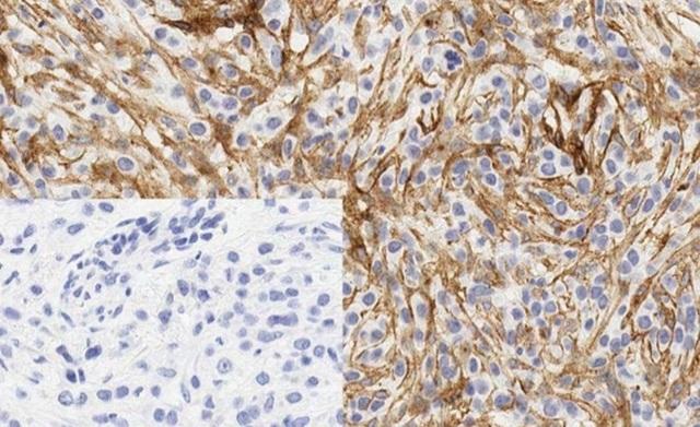
  • Stain 10 slides in under 2.5 hours using protocols pre-loaded on the BOND RX and BOND RXm instruments
  • Streamline your research needs with Abcam concentrated antibodies and BOND Titration Containers, kitted together

Learn More

Image
abcam-our-innovation-partners

技术参数

尺寸 & 重量

资源

BOND RTU 抗体和程序数据文件

这些数据文件可让您使用 BOND RX/RXm. 的最新程序版本。某些产品可能受当地许可限制;如有任何疑问,请联系当地的 Leica Biosystems 销售或支持代表。请确保下载正确的数据文件。

重要事项

  • 请不要将此更新应用于 BOND-III 或 BOND-MAX 系统。
  • 请下载适用于您所在地区的更新,并应用正确的更新。
  • 建议将所有 BOND 系统软件更新为最新版本,即 BOND RX 研究软件 v7.0
  • BOND RX 研究软件 v7.0 需要 BXD v25 或更高版本
  • 请不要将此更新应用于运行 RX/RXm v4.0 软件的系统;此文件不适用于软件 v4.0 的系统。

教育

从组织处理到数字病理,我们的解决方案为实验室提供高效、精准和可靠的支持。

Multiplex Fluorescence Immunohistochemistry using the Ultivue InSituPlex Platform on the Leica Biosystems BOND RX

Multiplex fluorescence immunohistochemistry offers a window into the biology of human disease, enabling the analysis of target protein expression in subsets of specific cells within the context of histopathological features of disease.