
Stereotaxic Accuracy

Humans and larger animals have great variation in brain size, and even in the relative size of different structures within brains of a given size. Fortunately for neuroscience research, rodent brains are made with more quality control; they are much more uniform animal to animal within a species and age range. This uniformity makes it possible to perform the same operation at the same location in several brains, but not in several others, and compare results. Published coordinate maps or atlases of the brain show the three-dimensional location of every structure in the young adult brain relative to a chosen zero point and plane. Such atlases exist for rat, mouse, hamster, and other small animal species. Bregma, a point on the skull where the bone plates have grown together, is usually used as the zero point. Together with a suitable manipulator and oriented head holder, such maps can be used to position probes at a preplanned site in the brains of several rodents. The instrument for moving a probe to a given coordinate in space – inside the brain – is a stereotaxic instrument.
Stereotaxic instrument function
The reason to place a probe at a specific location in brain is to study localization of function in the brain. The brain is a very heterogeneous structure, and different sites and regions have different functional roles to play. Learning to understand that organization is one of the major goals of neuroscience, and requires regional studies of brain. Various stains and immune-markers can show regional chemical variation in brain. Stereotaxic insertion of probes – for stimulation, injection, recording, microdialysis, lesion or other functions – is an important technique to gain understanding of brain functional organization.
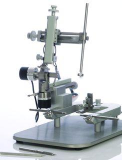
Traditional stereotaxic instruments consist of a head holder that is aligned with a manipulator, with the manipulator movements measured by vernier scales with a resolution of 100 microns. A newer idea is to use digital scales in place of vernier, and obtain easier, less error-prone reading and resolution to within 10 microns. Manipulators can be tilted and rotated, but doing so breaks the alignment with the head holder, and changes the distance that must be moved along each axis to reach the desired target. Sometimes an angled approach is necessary, to avoid arteries, or other structures important to the research. In this case, the investigator must perform math on the atlas coordinates, and usually pilot studies, to develop coordinates that work. Once that is done, the manipulator is locked in place, since further changes in tilt would again change the coordinates needed.
Straight up and down
Consequently, it is almost universal in stereotaxic research to do all surgeries for an experiment on several animals from the same angle of approach. This is usually straight down, 90 degrees from skull flat head alignment. This has been necessary given the instrumentation available, it is unfortunate because the resulting “action-at-target” variable is confounded with the “path to target” variable. Since they are linked, nothing distinguishes which one caused the change that is measured later.
Confounding
Imagine injecting deep in brain. Even with a glass pipette as a needle, some fluid will leak out the tip on the way down. When pressure is applied, some fluid will run up the side of the injection needle, where an opening is already made, rather than push back or flow into surrounding brain tissue. After an injection, when a pressurized bolus of fluid is at the tip, and the tip is withdrawn, the fluid will follow the needle back out. In the end, there is an intense injection at the target site, and a decreasingly intense injection along the needle track, usually all the way to the surface of the brain.
Later testing may show a behavioral change as a result of the injection, or uptake and transport to a remote site. But was that change due to the injection at the target site, or the injection somewhere higher up in the brain? Two variables that change in lock step are termed “Confounded,” and one cannot discern which was responsible for an observed result. A similar analysis applies with lesions, the electrode creates damage coming down, and always comes down through the same structures. Did the lesion damage at target cause the outcome alone, or in conjunction with the damage getting to the target?
Varying the angle of approach to avoid confounding
Suppose the same target was approached from a different angle, and through different brain structures, in several animals. In animals in which histology showed that the probe reached the correct target, the same result of the manipulation was seen. On control animals or on animals in which the target was not reached, the observed result was not obtained. This would be better science, better evidence that the “action at target” caused a certain outcome, than if the same result were obtained by all approaches from the same angle.
Technology
Based on the above considerations, NINDS funded an SBIR grant application to develop an instrument that would make it as easy to change angles as not to. This instrument has rotary encoders on the tilt and rotation movements, as well as linear encoders on the 3 linear axes, all of which are input into a computer. The computer can then keep track of where the probe tips is in space, and provide direction to reach the target, given the tilt angle and rotation angle.
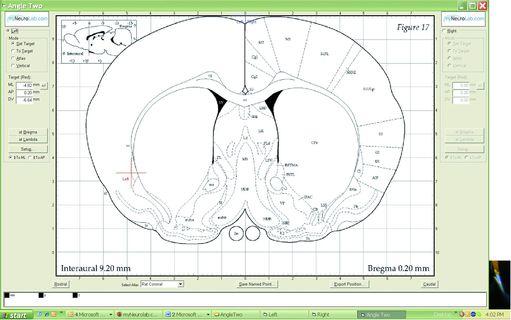
Experience a computer-guided stereotaxic demonstration
Dr. Charles Scouten presents a stereotaxic demonstration on Leica Angle Two
Read more about brain atlases
About the presenter
Charles Scouten acquired his Ph.D from SUNY Binghamton in 1980 with a dissertation entitled “Location of Neurons Mediating Androgen’s effect on Copulation, Urine Marking, and Body Weight”.
Related Content


Leica Biosystems Knowledge Pathway content is subject to the Leica Biosystems website terms of use, available at: Legal Notice. The content, including webinars, training presentations and related materials is intended to provide general information regarding particular subjects of interest to health care professionals and is not intended to be, and should not be construed as, medical, regulatory or legal advice. The views and opinions expressed in any third-party content reflect the personal views and opinions of the speaker(s)/author(s) and do not necessarily represent or reflect the views or opinions of Leica Biosystems, its employees or agents. Any links contained in the content which provides access to third party resources or content is provided for convenience only.
For the use of any product, the applicable product documentation, including information guides, inserts and operation manuals should be consulted.
Copyright © 2026 Leica Biosystems division of Leica Microsystems, Inc. and its Leica Biosystems affiliates. All rights reserved. LEICA and the Leica Logo are registered trademarks of Leica Microsystems IR GmbH.