
Neurotrauma from Impact

Brain damage from football, improvised explosive devices, and childhood falls has spurred a surge of funding for research on impact brain damage. Damage seems to result in slow growth of tangles of Tau proteins at the damaged sites, but behavioral changes due to the damage may not appear for years. This has delayed understanding of the causes of behavioral alterations now understood to be due to impact dementia.
Animal research is necessary to find any early indicators of impact and to test therapies. One preliminary finding has been that a repeated impact before the first damaged tissue has healed and stabilized results in a much worse outcome than one impact alone. This has implications for how a concussion should be treated. The patient, although feeling fine and fully recovered, should not go back to the battlefield, or the football field, for a considerable time that needs to be defined.
Several instruments have been developed to create controlled impacts on the skull or brain, or spinal cord, of research animals. There are pneumatic, gravity and electrically driven devices. Prior to 2006, such instruments were massive, noisy, and awkward to position.
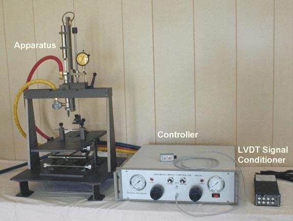
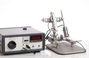
In 2006, a device came on the market with a small, lightweight impeller, and the entire actuator could be mounted on a stereotaxic instrument. Mounting on a stereotaxic instrument allows positioning the position and angle of the impact reproducibly relative to Bregma with 10 micron resolution. Depth of penetration is also set with the stereotaxic instrument, with the aid of a contact sensor. Impacts may be either open head or closed head, that is, directly on the brain, or on the skull surface. Both have been shown to generate permanent neural damage.
How do you select what instrument is best for your experiments?
The first question usually asked is “how much force does it have?” This is the wrong question, whose answer, at least with open head injury, for all of these devices, is zero, or very nearly so. Simply put, the brain does not push back. A force sensor between the brain and the Impactor tip would read very little. Further, with a slow push, the brain will compress and reposition and avoid much damage, if the entry is not too deep or extreme. So depth is not entirely the right measure. So what is the relevant variable?
Momentum might be, if the probe was going to be stopped by the target. However, these devices have preset stops that limit penetration. The moving part is stopped by a metal stop, not the target tissue. If two identical probes, but one with a ton behind it, and one with an ounce behind it, traveling at the same velocity, impact the brain, are not slowed by it, and penetrate to a depth where it is then stopped by its own stop, the result should be identical. Except stopping the ton probe will require great force, and it will probably overshoot, and then vibrate in the brain much more than the ounce probe, and make a very loud impact noise.
Further, there is always a limited distance available for acceleration, and there must be very fast acceleration to work up to a reasonable impact velocity. More mass will require greater force to be applied during acceleration, which will require more power, a more expensive controller, and more noise to stop it.
So in the end, a light impeller probe with controlled velocity and preset maximum depth of penetration should yield more reproducible results. The probe must have enough mass that it is not stopped or slowed noticeably by the brain tissue. That requirement is easy to meet if the mass is moving at high velocity.
Some contend that the duration the probe is in contact is crucial, others say not. Most instruments can control the contact duration, and that will remain until rigorous tests show duration does or does not affect the degree of damage.
About the presenter
Charles Scouten acquired his Ph.D from SUNY Binghamton in 1980 with a dissertation entitled “Location of Neurons Mediating Androgen’s effect on Copulation, Urine Marking, and Body Weight”.
Related Content


Leica Biosystems Knowledge Pathway content is subject to the Leica Biosystems website terms of use, available at: Legal Notice. The content, including webinars, training presentations and related materials is intended to provide general information regarding particular subjects of interest to health care professionals and is not intended to be, and should not be construed as, medical, regulatory or legal advice. The views and opinions expressed in any third-party content reflect the personal views and opinions of the speaker(s)/author(s) and do not necessarily represent or reflect the views or opinions of Leica Biosystems, its employees or agents. Any links contained in the content which provides access to third party resources or content is provided for convenience only.
For the use of any product, the applicable product documentation, including information guides, inserts and operation manuals should be consulted.
Copyright © 2026 Leica Biosystems division of Leica Microsystems, Inc. and its Leica Biosystems affiliates. All rights reserved. LEICA and the Leica Logo are registered trademarks of Leica Microsystems IR GmbH.