




Digital-Ready Slides
Updated Date: 7 August 2026
Glass: The Clear Choice for AI-Ready Digital Pathology
As digital pathology adoption grows, glass coverslips help deliver the clarity and consistency needed for efficient workflows and advanced tools, making them the trusted choice today and for future AI-ready applications.
When digital imaging and AI analysis depend on every pixel, “good enough” risks missed details and wasted time.
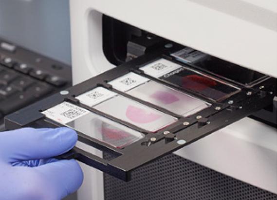
Glass
|
Film
|
|
Highly Stable and Uniform SurfaceMaintains a smooth profile, crucial for consistent and reliable digital focusing. |
Irregular and Rippled SurfaceShows height variations that compromise image consistency and quality. |
|
Image

Optimized for AI and EfficiencyCleaner scans reduce the need for rescans, saving time and enabling accurate AI analysis |
Image

Risk of Inefficient WorkflowsCan lead to suboptimal scans, reducing lab efficiency and requiring time-consuming rescans. |
Why Glass
|
Study Highlights
|


The Leica Biosystems Advantage: Designed for Digital Readiness
|
|
Snowcoat Slides |
|
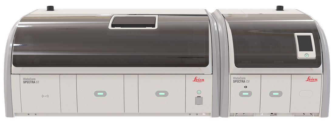
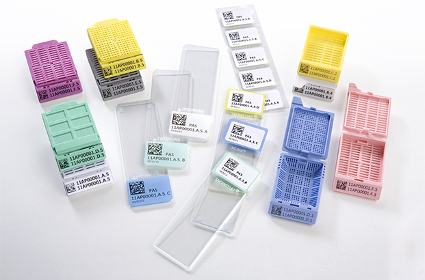
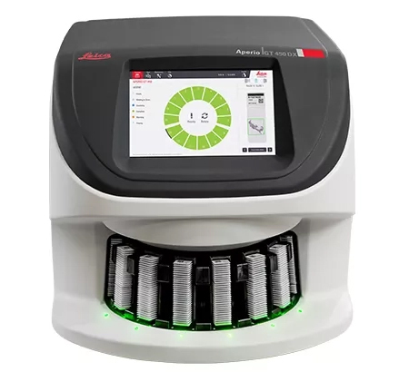
Our expertise in pathology workflows enables us to deliver integrated solutions - including digital-ready glass coverslips - that streamline operations today and position laboratories for a digital, AI-powered future.
* Study on the performance of different cover glasses in microscopy, conducted by a renowned institute in Jena, Germany. Commissioned by Leica Biosystems GmbH, Nussloch, Germany; September 2025. Sample size: 6 total samples (3 with cover glass, 3 with cover film).
For In Vitro Diagnostic Use
Glass vs Film FAQs
Why does coverslip material matter in digital pathology?
Coverslip material can influence image consistency, optical clarity, and scan quality in digital pathology workflows. Surface smoothness and optical stability are important considerations for whole-slide imaging and AI-enabled pathology applications.
What did the glass vs. film study evaluate?
The study evaluated the surface characteristics, optical properties, and image quality factors associated with glass and polymer film coverslips in digital pathology workflows, including surface roughness, waviness, optical transmission, and defect rates. *
What were the key findings of the study?
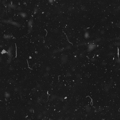
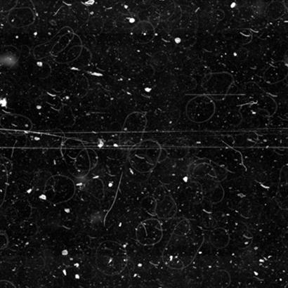
The study observed clear differences between glass and polymer film coverslips. Film-covered samples exhibited approximately five times more surface defects compared to glass-covered samples, along with visible longitudinal scratches that were not observed on glass-covered samples. *
The study also observed differences in surface texture, flatness, and optical consistency that may influence image quality and rescanning rates in digital pathology workflows.
Why is surface flatness important in whole-slide imaging?
Surface flatness helps support consistent digital focusing during whole-slide scanning and may reduce focus variation, image inconsistency, and optical artifacts.
The study found that glass coverslips maintained smoother and more uniform surface profiles, while film samples exhibited irregular corrugations and rippled surface structures. *
Can surface imperfections impact digital scans?
Yes. Surface imperfections such as scratches, roughness, waviness, or contamination may contribute to visual artifacts that can affect scan quality and image consistency.
The study observed approximately five times more surface defects on film-covered samples compared to glass-covered samples. *
How can coverslip quality influence rescanning rates?
Image inconsistencies, surface artifacts, and optical irregularities may contribute to rescans and workflow interruptions in high-volume digital pathology environments.
The study observed local image quality degradation and reduced contrast in film-covered samples, which may impact scanning consistency. *
Why is image consistency important for AI-enabled pathology workflows?
AI-assisted image analysis relies on consistent, high-quality digital images. Variability in slide presentation, image contrast, or scan quality may affect workflow efficiency and image interpretation consistency.
As digital pathology adoption expands, image consistency is becoming increasingly important for AI-enabled workflows and large-scale image analysis applications.
Does the study suggest that glass coverslips better support digital pathology workflows?
The study findings suggest that glass coverslips may better support digital pathology workflows by providing smoother surface characteristics, fewer defects, and more consistent imaging conditions for whole-slide scanning. *
What did the study observe regarding image quality?
Optical simulations within the study showed minimal optical performance impact for glass coverslips, while film-covered samples demonstrated localized reductions in image contrast and resolution under certain conditions. *
The study also observed that film surfaces exhibited irregular corrugations and greater micro roughness compared to glass. *
What are common considerations when evaluating glass vs. film workflows?
Laboratories may evaluate image consistency, surface quality, rescanning rates, throughput requirements, workflow efficiency, and compatibility with digital pathology and AI-enabled workflows.
What is the main takeaway from the glass vs film discussion?
As digital pathology adoption continues to grow, image consistency and scan quality are becoming increasingly important workflow considerations.
The study findings suggest that glass coverslips may provide advantages in digital pathology workflows by delivering smoother surfaces, fewer defects, and more consistent imaging conditions for whole-slide scanning and AI-enabled applications. *
* Study on the performance of different cover glasses in microscopy, conducted by a renowned institute in Jena, Germany. Commissioned by Leica Biosystems GmbH, Nussloch, Germany; September 2025. Sample size: 6 total samples (3 with cover glass, 3 with cover film).
Related Content

Leica Biosystems content is subject to the Leica Biosystems website terms of use, available at: Legal Notice. The content, including webinars, training presentations and related materials is intended to provide general information regarding particular subjects of interest to health care professionals and is not intended to be, and should not be construed as, medical, regulatory or legal advice. The views and opinions expressed in any third-party content reflect the personal views and opinions of the speaker(s)/author(s) and do not necessarily represent or reflect the views or opinions of Leica Biosystems, its employees or agents. Any links contained in the content which provides access to third party resources or content is provided for convenience only.
For the use of any product, the applicable product documentation, including information guides, inserts and operation manuals should be consulted.
Copyright © 2026 Leica Biosystems division of Leica Microsystems, Inc. and its Leica Biosystems affiliates. All rights reserved. LEICA and the Leica Logo are registered trademarks of Leica Microsystems IR GmbH.