Sacrifice Perfusion System Controlled Pressure
The Perfusion Two Automated Pressure Perfusion system includes an automated air compressor that quietly pumps the air tank of the Perfusion One up to 300 mm Hg and stops. As each animal is being prepared, the pressure is set up to begin at the turn of a switch.
Pressure above physiological pressure is required to clear all blood vessels and leave no red blood cells. Automating the perfusion system gives much greater consistency and more reproducable perfusions, as well as greater convenience and ease of operation.
Learn more
Quicker Fixation
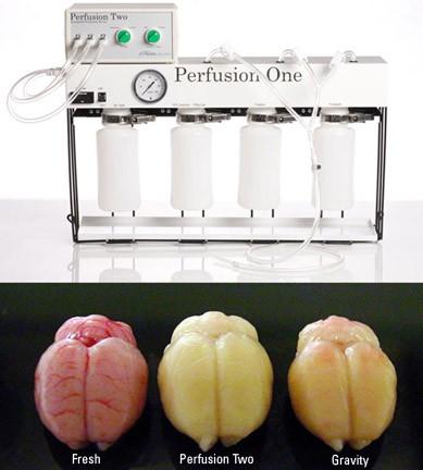
Pressure washout gets all blood cells out in about 5 seconds. Fixative can get in sooner.
Better HRP or Fluorescence Results
Removing all red bloods cells reduces HRP background and autofluoroesence
Better Tissue Quality
Any red blood cells left block capillaries, and thus access of fixative to tissue downstream. Fast removal of all of them yields more even fixation.
Avoid Soft Tissue Shrinkage
With proper management of fixative tonicity, discussed in the references, you can avoid tissue shrinkage.
Testimonials
"Bronson Methodist Hospital: a systematic approach to validating xylene‑free processing"

Leica PELORIS improving workflow, safety, and productivity

Excellent results with Leica PELORIS xylene-free processing

Going green with a next generation tissue processor

Publications
The following publications cite the Leica Biosystem’s Perfusion One/Two for Research Use Only.
- Kirichenko, E. Y., Matsionis, A. E., Povilaitite, P. E., Akimenko, M. A., & Logvinov, A. K. (2017). Characteristics of the Structural Organization of the Ventral Posteromedial and Posterolateral Nuclei and the Reticular Nucleus of the Thalamus in Rats (an immunohistochemical study). Neuroscience and Behavioral Physiology, 47(6), 621–626. doi.org/10.1007/s11055-017-0444-9
- Berger, C., Kühne, D., Scheper, V., & Kral, A. (2017). Congenital deafness affects deep layers in primary and secondary auditory cortex. Journal of Comparative Neurology, 525(14), 3110–3125. doi.org/10.1002/cne.24267
- Norcini, M., Sideris, A., Adler, S. M., Hernandez, L. A. M., Zhang, J., Blanck, T. J. J., & Recio-Pinto, E. (2016). NR2B Expression in Rat DRG Is Differentially Regulated Following Peripheral Nerve Injuries That Lead to Transient or Sustained Stimuli-Evoked Hypersensitivity. Frontiers in Molecular Neuroscience, 9, 100. doi.org/10.3389/fnmol.2016.00100
- Pattwell, S. S., Liston, C., Jing, D., Ninan, I., Yang, R. R., Witztum, J., … Lee, F. S. (2016). ARTICLE Dynamic changes in neural circuitry during adolescence are associated with persistent attenuation of fear memories. Nature Communications, 7. doi.org/10.1038/ncomms11475
- Dincheva, I., Drysdale, A. T., Hartley, C. A., Johnson, D. C., Jing, D., King, E. C., … Lee, F. S. (2015). FAAH genetic variation enhances fronto-amygdala function in mouse and human HHS Public Access. Nat Commun. doi.org/10.1038/ncomms7395
- Richards, K., Calamante, F., Tournier, J.-D., Kurniawan, N. D., Sadeghian, F., Retchford, A. R., … Petrou, S. (2014). Mapping somatosensory connectivity in adult mice using diffusion MRI tractography and super-resolution track density imaging. NeuroImage, 102 Pt 2, 381–392. doi.org/10.1016/j.neuroimage.2014.07.048
- Tapia, J. C., Kasthuri, N., Hayworth, K., Schalek, R., Lichtman, J. W., Smith, S. J., & Buchanan, J. (2013). High contrast en bloc staining of neuronal tissue for field emission scanning electron microscopy. Nature Protocols, 7(2), 193–206. doi.org/10.1038/nprot.2011.439
- Kim, T. H., Richards, K., Heng, J., Petrou, S., & Reid, C. A. (2013). Two lines of transgenic mice expressing cre-recombinase exhibit increased seizure susceptibility. Epilepsy Research, 104(1), 11–16. doi.org/10.1016/j.eplepsyres.2012.10.005
- Deane, J. A., Verghese, E., Martelotto, L. G., Cain, J. E., Galtseva, A., Rosenblum, N. D., … Ricardo, S. D. (2013). Visualizing renal primary cilia. Nephrology, 18(3), 161–168. doi.org/10.1111/nep.12022