Chromogenic Stain of Human Tissue with ChromoPlex III and BOND RX

ChromoPlex III Triple Detection Chomogenic Multiplex Kit for the BOND RX/RXm Automated Stainers

Simplify chromogenic multiplex IHC & ISH with a plug-and-play detection kit designed for simplicity, consistency, and seamless integration into digital workflows.

Plug-and-Play Simplicity. Designed for Digital.

ChromoPlex III is a three-plex chromogenic detection kit for the BOND RX and RXm automated stainers that simplifies assay setup and staining. Researchers can run up to three markers in a single tissue section. Pick your antibodies, assign to colors (brown, red, or green), load on BOND RX stainer, and start your run.

Image
Illustration of Chromogenic Stain of Human Tissue with ChromoPlex III and Digital Pathology Image Analysis
Image
ChromoPlex III Multiplex Chromogenic Detection Kit

Plug-and-Play Multiplexing

Detection reagents come in one simple kit. No need for conjugating antibodies.

Image
Built for Consistency

Built for Consistency

Pre-optimized reagents and protocols on the BOND research platform to reduce run-to-run and user-to-user variability. Uses trusted BOND detection chemistry.

Image
Chromogenic Stain Image Analysis of Human Tissue with ChromoPlex III Detection Kit

Designed for Image Analysis

Crisp and clear staining without signal amplification—which has shown to preserve linearity between antigen expression and signal intensity for biomarker quantification.

How it Works: Triple-Layer Sequential Chromogenic Staining

Why this matters:

  • Each chromogen is applied after a fresh elution step.
  • Tissue integrity and morphology are preserved.
  • Chromogen signals remain clear and non-overlapping.
  • Ideal for cell-level and spatial quantification.
Image
Triple-layer Sequential Chromogenic Staining using BOND-RX and ChromoPlex III

Proven Performance on Tumor Samples

Triplex Characterization of Breast & GI Cancers

Using ChromoPlex III, researchers stained HER2 / PD-L1 / CD3 and HER2 / PD-L1 / CD8 panels on FFPE breast and GI carcinomas, revealing:

  • Distinct variation in TIL (CD3+, CD8+) infiltration.
  • PD-L1 expression heterogeneity.
  • Clustering of T cells near HER2+ tumor cells.
  • Clear spatial interactions quantifiable by image analysis.

Peak immune-tumor proximity occurred at ~10uM, highlighting the power of chromogenic multiplexing in spatial biology.

Image
ChromoPlex III stains of Breast and Colon Carcinomas
Figure 1: Multiplex IHC staining of Breast and Colon Carcinoma: CD3-DAB, HER2(BMA)-Red, PDL1-Green shown in b, d, f, h and CD8-DAB, HER2(BMA)-Red, PDL1-Green shown in (a, c, e, g). Staining exhibits the spatial interaction of TILs with the carcinoma. Sample images are shown demonstrating staining of two 3Plex panels on 4 representative cases, 2 colon carcinoma (c, d and g, h) and 2 breast carcinoma (a, b and e, f). Images shown at 20x slides scanned on Leica GT450 Dx.
Image
Image analysis using HALO
Figure 2: Image analysis performed using HALO digital image analysis software. Breast carcinoma tissue stained with HER2 (BMA), CD8 and PD-L1 (CAL10) visualized with ChromoPlex III Triple Detection RUO (a). Tissue was analyzed with HALO AI to segment sells based off nuclear and membrane morphology (b). Biomarker positivity was measured with HALO’s Multiplex IHC analysis module. Cellular compartment positivity is indicated with the colored overlay. HALO proximity analysis measured the distance of each CD8 positive cell to the nearest HER2 positive tumor cell (c). CD8+ cells are listed according to their distance away from HER2+ cells and show a peak at 10 µm (d). Scale bar is 50 µm. Stomach carcinoma tissue stained with HER2 (BMA), CD3 and PD-L1 (CAL10) visualized with ChromoPlex III Triple Detection RUO (e). Tissue was analyzed with HALO AI to segment sells based off nuclear and membrane morphology (f). Biomarker positivity was measured with HALO’s Multiplex IHC analysis module. Cellular compartment positivity is indicated with the colored overlay. HALO proximity analysis measured the distance of each CD3 positive cell to the nearest HER2 positive tumor cell (g). CD3+ cells are listed according to their distance away from HER2+ cells and show a peak at 10 µm (h). Scale bar is 50 µm.

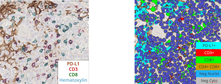
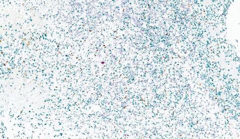
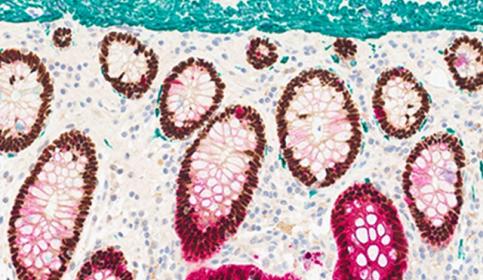
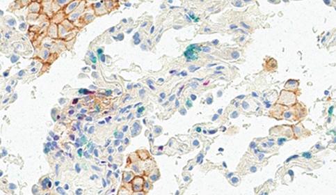
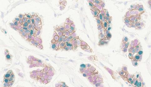
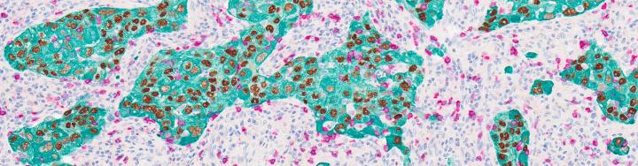
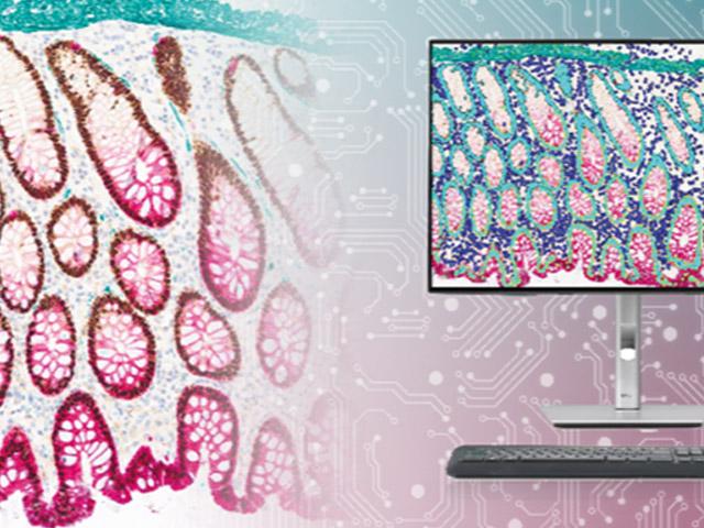
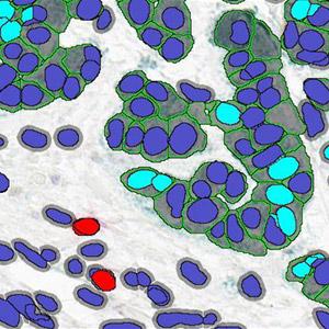
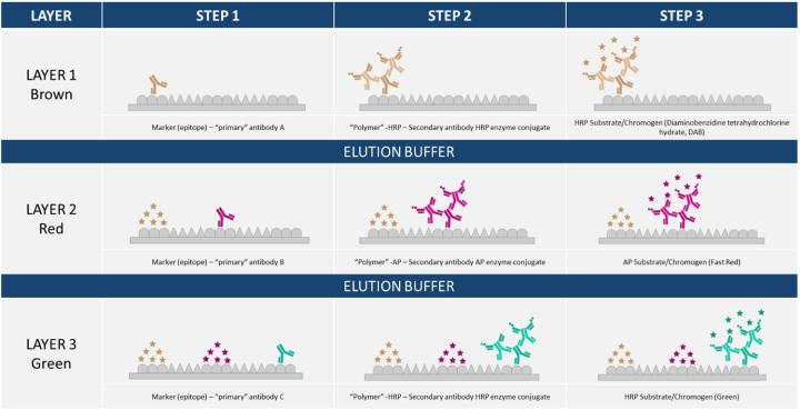
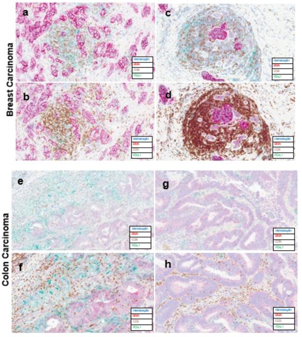
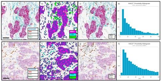
Built for Digital Pathology and Image Analysis

ChromoPlex III detection delivers crisp chromogenic signal without amplification, which:

  • Preserves linearity between antigen load and stain intensity.
  • Improves feature recognition for AI segmentation.
  • Supports accurate cell classification across compartments.
  • Integrates seamlessly with the Aperio GT 450 scanner and HALO.
Image
Chromogenic Signal

Plug-and-play Multiplexing on BOND RX Stainer

Discover a streamlined workflow that lets you focus on discovery, not optimization:

  1. Choose your three primary markers.
  2. Assign chromogens.
  3. Load reagents and slides into BOND RX stainer.
  4. Start your automated multiplex run.
  5. Scan with Aperio GT 450 scanner or FL 120 scanner.
  6. Quantify with HALO or HALO AI.

The BOND RX stainer balances a researcher’s need for customization with the translational need for standardization.

Image
BOND Rx

Compatible with Thousands of Validated Antibodies

Leica Biosystems and Abcam provide one of the world’s largest portfolios of IHC-validated antibodies.

  • Abcam’s portfolio includes 13,000 recombinant antibodies with 6,300 validated on the BOND RX scanner and bundled antibody kits with BOND Titration Containers available.

Bring your own primaries or choose from Leica Biosystems’ catalog of RUO antibodies.

Image
abcam official black and white logo with slogan progress happens together

Customer Spotlight: Dana-Farber Validation

Namrata Singh, PhD

Technical Director, Research Scientist – Dana-Farber Cancer Institute

“Today I successfully validated ChromoPlex III on the BOND RX on ovarian carcinoma and GBM using p53, Ki-67, and PD-L1. The results showed outstanding staining quality, specificity, and reproducibility.

These findings highlight how advanced chromogenic multiplexing can be seamlessly integrated into automated platforms like the BOND RX, enabling deeper spatial and phenotypic insights into the tumor microenvironment”

Image
Chromoplex III Tissue from Dana Farber
Image
Chromoplex III Tissue from Dana Farber
Image
Chromoplex III Tissue from Dana Farber

ChromoPlex III Interactive Digital Slide Gallery

Explore whole slide images scanned on the Aperio GT 450 slide scanner. Change magnification, navigate, and analyze biomarker patterns in high resolution using our digital slide viewer.

Image
Chromogenic Stain of Colon Tissue with ChromoPlex III and BOND RX

Tissue: Colon

CDX2 | CK20 | Des

View slide

Image
Chromogenic Stain of Lung Tissue with ChromoPlex III and BOND RX

Tissue: Lung

PD-L1 [CAL10] | FOXP3 | CD8

View slide

Image
Chromogenic Stain of Breast Tissue with ChromoPlex III and BOND RX

Tissue: Breast

HER2 [BMA] | ER | PR

View slide

Leica Biosystems’ End-to-End Portfolio for Translational Research

ChromoPlex III integrates seamlessly across Leica Biosystems’ staining, scanning, and Indica Lab’s image management and analysis platforms—streamlining today’s workflow and enabling tomorrow’s breakthroughs.

Image
Leica Biosystems Chromogenic Staining Workflow Solutions for Translational Research

Experience the simplicity and integration of ChromoPlex III

Talk to an expert

FOR RESEARCH USE ONLY. Not For Use In Diagnostic Procedures