
Eliminating Rework Through Utilization of Interfacing Automated Staining Platforms
Many pathology laboratories face similar issues in today’s healthcare environment: handling higher volumes of blocks and slides with fewer experienced histotechnologists. Many laboratories turn to lean assessments to help identify wastes in their current processes. The goal of these assessments is to reduce those identified wastes to help increase the productivity of the available histotechnologists. One of the more common wastes found in the histology laboratory is rework or duplicate data entry into automated staining platforms.
Method
The Leica Biosystems Customer Education Team had the opportunity to partner with an Anatomic Pathology laboratory to investigate the benefit of an interface for an Immunohistochemical staining instrument. This laboratory uses multiple Laboratory Information Systems (LIS), but only one system interfaces with the IHC stainer. An order placed in the interfaced LIS automatically transfers to the IHC staining instrument. The histotechnologist can easily search through the IHC stainer controller, find the stain order for the specific slide, and print the label. For the non-interfaced LIS, after placing the order in the LIS, the histotechnologist must enter the case accession number, block ID, and patient name and select the stain to be run manually. After entering the information into the IHC stainer controller, the histotechnologist can print the label. During a multiple-day observation period, the Leica Biosystems Customer Education Team performed time and motion studies to collect data for each LIS process. The goal was to provide a side-by-side comparison of the required time to prepare staining labels for the interfaced LIS and the non-interfaced LIS.
Results
Discussion
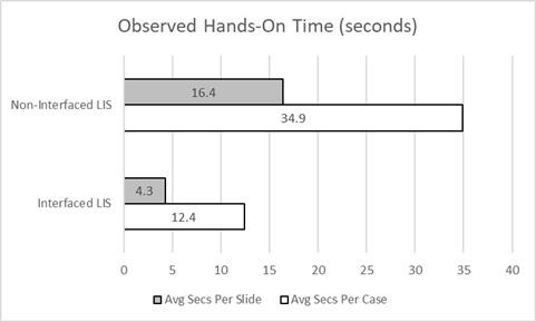
The chart shows both times, in seconds, by case, and by slide, to offer an accurate comparison. The manual process is faster when entering multiple stain orders for specific cases since not all information needs to be entered for each stain. The case accession number, patient name, and sometimes the block ID could remain the same for multiple stain entries. The average number of stains per case was 2.4 for the observation period at this laboratory.
Analyzing the average differences in time by stain, or slide, for the interfaced and the non-interfaced LIS, the team found that, for this laboratory, the histotechnologists spend 3.8 times longer preparing the staining labels for non-interfaced LIS stain orders as they do for interfaced LIS stain orders. This laboratory performs 48,000 IHC slides annually with the non-interfaced LIS; that time difference equates to 161.3 hours of histotechnologists time. That means the histotechnologists spend over one month annually preparing the staining labels.
Conclusion
For this Anatomic Pathology laboratory and similar laboratories not utilizing interfaces for their staining platforms, there are time savings available by removing rework and duplicate data entry. Although this study focused on time savings, anytime data entry steps are added to a process; there is an inherent risk of data entry errors. Eliminating those possible errors is another benefit of utilizing LIS interfaces for automated staining platforms.
Projections and Realized Results are specific to the institution where they were obtained and may not reflect the results achievable at other institutions.
About the presenter
David Newell has been working in the field of Laboratory Medicine for over 20 years. He has experience working on the Laboratory Informatics Implementation team, leading installations across North America. Before joining Leica Biosystems, he served in several roles at a large reference laboratory in Arizona, including over eight years as a Histotechnologist. He has experience managing an Anatomic Pathology Department for a level 1 trauma center and a dermatology laboratory. David holds a Bachelor of Science in Kinesiology and Master of Business Administration and Histologic Technician (HT) certification through the American Society for Clinical Pathology (ASCP). He received his Six Sigma and Lean training through Quest Diagnostics in 2002 and 2010, respectively.
Related Content


Leica Biosystems Knowledge Pathway content is subject to the Leica Biosystems website terms of use, available at: Legal Notice. The content, including webinars, training presentations and related materials is intended to provide general information regarding particular subjects of interest to health care professionals and is not intended to be, and should not be construed as, medical, regulatory or legal advice. The views and opinions expressed in any third-party content reflect the personal views and opinions of the speaker(s)/author(s) and do not necessarily represent or reflect the views or opinions of Leica Biosystems, its employees or agents. Any links contained in the content which provides access to third party resources or content is provided for convenience only.
For the use of any product, the applicable product documentation, including information guides, inserts and operation manuals should be consulted.
Copyright © 2026 Leica Biosystems division of Leica Microsystems, Inc. and its Leica Biosystems affiliates. All rights reserved. LEICA and the Leica Logo are registered trademarks of Leica Microsystems IR GmbH.