SlideQC BF
Developed by Indica Labs, SlideQC BF is a deep learning tool for images that enables an automated and robust quality control workflow by detecting the most common artifacts generated during slide preparation and digital scanning. Artifacts include air bubbles, dust/debris, tissue folds, out-of-focus areas, and pen marks on H&E and immunohistochemistry (IHC) stained slides.
SlideQC BF is For Research Use Only and not intended for clinical diagnostic use. SlideQC BF accessed via the Aperio HALO AP image management system.

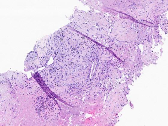
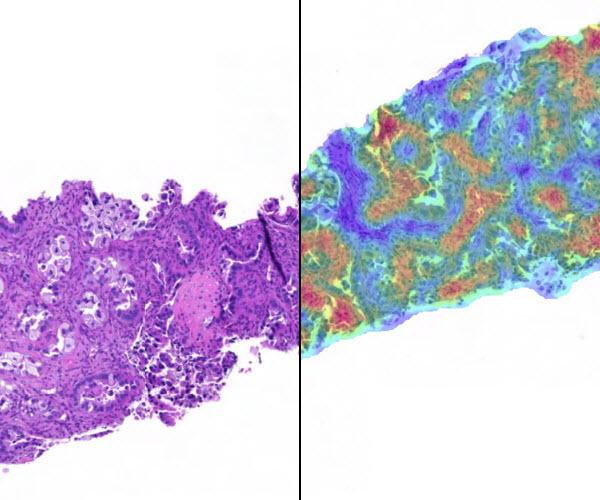
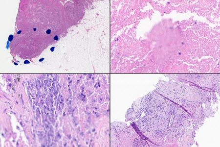
Step 1: H&E or IHC image of any tissue type
SlideQC BF robustly and automatically detects the most common artifacts found in digitized samples and brings efficiency to the quality control workflow.
The algorithm can be launched by a user directly in the viewer or configured to automatically run in Aperio HALO AP.
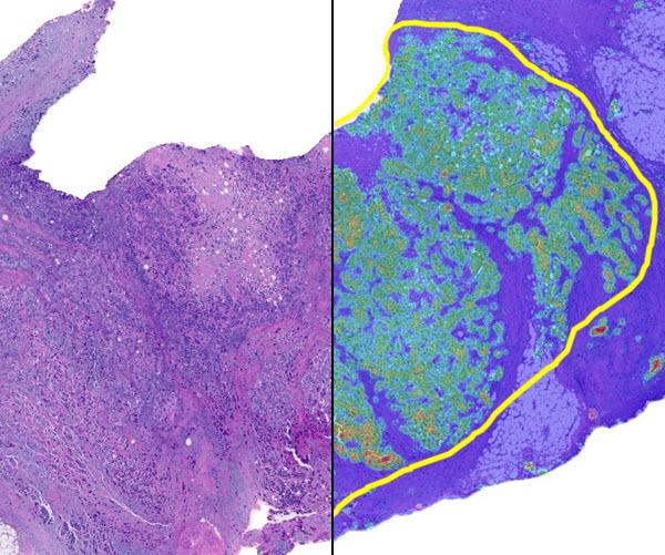
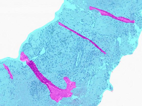
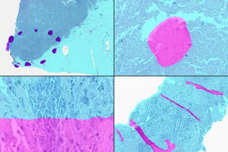
Step 2: Detection of Artifacts
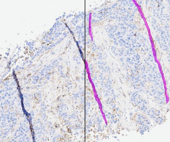
SlideQC BF generates an image mask across the entire WSI classifying artifact such as air bubbles, folds, dust/debris, out of focus regions and pen mark.
- Acceptable Tissue: Cyan
- Artifact: Pink
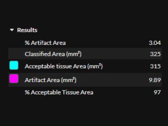
Results: Quantitative
SlideQC BF AI can generate an automated and customizable tag flagging slides which exceed a user defined % artifact threshold. The following metrics as output for across the mage:
- Artifact Area (%)
- Classified Area (mm²)
- Acceptable Tissue Area (mm²)
- Artifact Area (mm²)
- Acceptable Tissue Area (%)
SlideQC BF in Aperio HALO AP Demonstration
Explore the future of pathology with the SlideQC BF app from Indica Labs. SlideQC BF seamlessly integrates within the Aperio HALO AP enterprise digital pathology platform to enable an automated and robust quality control workflow. This app detects the most common artifacts generated during the process of slide preparation and scanning including air bubbles, dust/debris, folds, out-of-focus areas, and pen marks.
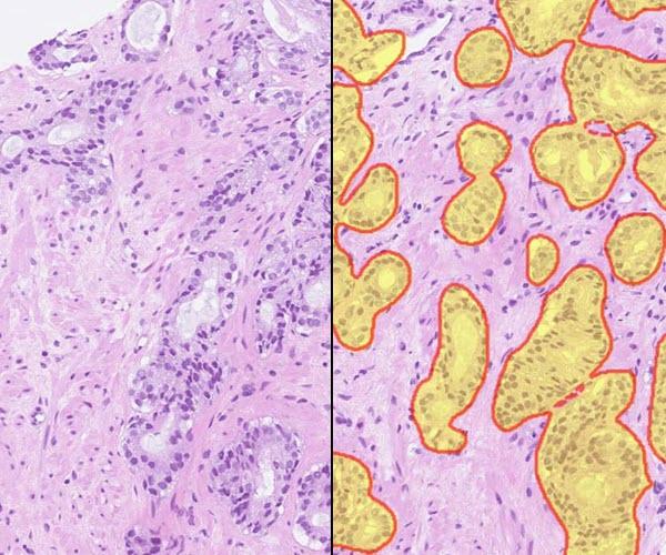
After selecting an image, view the H&E or IHC slide in the viewer and the SlideQC BF results. Results include overlays and quantitative results. The overlay highlights artifacts in magenta and acceptable tissue areas in cyan.
Use the quantitative SlideQC BF results to automatically tag images exceeding a laboratory-defined artifact threshold for recut or rescan. These images can be triaged into separate worklists for additional work.
Artifacts in Digital Pathology
High quality slides are a prerequisite for a first-rate digital workflow. Artifacts generated in the slide creation and scanning process can not only affect interpretation but can prevent image analysis and AI tools from rendering the correct results. SlideQC BF automatically detects and annotates artifacts present on images, increasing the accuracy and quality of the analysis results while maximizing scanned tissue. Artifacts may include pen marks, out of focus areas, tissue folds, debris, or bubbles.
Related Product Applications