
Aperio GT 450 Improves Throughput by 64% and Reduces QC Tech Time by 94% in Workflow Study


NeoGenomics is a large research and reference laboratory located in California and has experienced significant increases in annual slide volumes. The NeoGenomics senior management team was tasked with evaluating ways to scale up digital pathology operations to keep up with increasing demand. Consequently, the laboratory director desired to perform a comparative research study to assess the performance of the Aperio GT 450 versus Aperio AT2. Throughput, QC tech time and other performance metrics were measured.
Methodology
Samples
A total of 155 slides, that were representative samples from the high volume reference laboratory, were part of this study. Samples included:
- 35 individual H&E stained slides
- 36 breast IHC slides evaluated for key biomarkers along with 10 associated breast H&E slides
- 26 colon IHC stained slides for key biomarkers along with 8 associated colon H&E slides
- 33 squamous mucosa CISH stained slides for HPV biomarkers (RNA ISH) along with 7 associated H&E slides
H&E stained slides
35 Individual H&E stained slides included in the study
| Organ | Method | # of slides |
|---|---|---|
| Brain | H&E | 1 |
| Cervix | H&E | 2 |
| Gall bladder | H&E | 1 |
| Lung | H&E | 4 |
| Prostate | H&E | 20 |
| Right iliac bone | H&E | 1 |
| Soft palate | H&E | 1 |
| Stomach | H&E | 3 |
| Tongue | H&E | 1 |
| Uterus | H&E | 1 |
| Total | 35 |
IHC breast biomarkers
36 breast IHC slides evaluated for key biomarkers along with 10 associated breast H&E slides were included in the study.
| Organ | Method | Stain | # of slides | Case # |
|---|---|---|---|---|
| Breast | IHC/H&E | HER2, ER, PR, Ki67, H&E | 5 | 1 |
| Breast | IHC/H&E | HER2, ER, PR, Ki67, p53, H&E | 5 | 2 |
| Breast | IHC/H&E | ER, PR, Ki67, p53, H&E | 5 | 3 |
| Breast | IHC/H&E | ER, PR, Ki67, p53, H& | 5 | 4 |
| Breast | IHC/H&E | ER, PR, Ki67, p53, H&E | 5 | 5 |
| Breast | IHC/H&E | HER2, ER, PR, Ki67, H&E | 5 | 6 |
| Breast | IHC/H&E | HER2, PR, KI67, H&E | 4 | 7 |
| Breast | IHC/H&E | HER2, Ki67, p53, H&E | 4 | 8 |
| Breast | IHC/H&E | HER2, Ki67, p53, H&E | 4 | 9 |
| Breast | IHC/H&E | HER2, Ki67, p53, H&E | 4 | 10 |
| Total slides | 46 |
Colon biomarkers
26 colon IHC stained slides evaluated for key biomarkers along with 8 associated colon H&E slides were included in the study.
| Organ | Method | Stain | # of slides | Case # |
|---|---|---|---|---|
| Colon | IHC/H&E | MLH-1, MSH-2, MSH-6, PMS-2, H&E | 5 | 1 |
| Colon | IHC/H&E | MLH-1, MSH-2, MSH-6, PMS-2, H&E | 5 | 2 |
| Colon | IHC/H&E | MLH-1, MSH-2, MSH-6, PMS-2, H&E | 5 | 3 |
| Colon | IHC/H&E | MLH-1, MSH-2, MSH-6, PMS-2, H&E | 5 | 4 |
| Colon | IHC/H&E | MLH-1, MSH-2, MSH-6, PMS-2, H&E | 5 | 5 |
| Colon | IHC/H&E | MSH-2, MSH-6, H&E | 3 | 6 |
| Colon | IHC/H&E | MLH-1, PMS-2, H&E | 3 | 7 |
| Colon | IHC/H&E | MLH-1, PMS-2, H&E | 3 | 8 |
| Total slides | 34 |
Squamous mucosa RNA ISH biomarkers
33 squamous mucosa CISH stained slides evaluated for HPV biomarkers (RNA ISH) along with 7 associated H&E slides were included in the study.
| Organ | Method | Stain | # of slides | Case # |
|---|---|---|---|---|
| Squamous Mucosa | CISH/H&E | HPV 16/18, HR 18, HPV LR10, HPV RNA-, HPV RNA+, HPV H&E | 6 | 1 |
| Squamous Mucosa | CISH/H&E | HPV 16/18, HR 18, HPV LR10, HPV RNA-, HPV RNA+, HPV H&E | 6 | 2 |
| Squamous Mucosa | CISH/H&E | HPV 16/18, HR 18, HPV LR10, HPV RNA-, HPV RNA+, HPV H&E | 6 | 3 |
| Squamous Mucosa | CISH/H&E | HPV 16/18, HR 18, HPV LR10, HPV RNA-, HPV RNA+, HPV H&E | 6 | 4 |
| Squamous Mucosa | CISH/H&E | HPV 16/18, HR 18, HPV LR10, HPV RNA-, HPV RNA+, HPV H&E | 6 | 5 |
| Squamous Mucosa | CISH/H&E | HPV 16/18, HR 18, HPV LR10, HPV RNA-, HPV RNA+, HPV H&E | 5 | 6 |
| Squamous Mucosa | CISH/H&E | HPV 16/18, HR 18, HPV LR10, HPV RNA-, HPV RNA+, HPV H&E | 5 | 7 |
| Total slides | 40 |
Equipment
| Equipment Type / Description | Identification / Serial Number |
|---|---|
| Aperio GT 450 next generation scanner | Serial number 12001 with hardware version 1.0.1 and controller version 1.0.0.5055 |
| Dell R740XL DSR server loaded with Aperio eSlide Manager and Aperio ImageScope Version 12.4. | Aperio eSlide Manager Version 12.4.2.5010 |
| Dell 5820 workstation with ImageScope version 12.4, Win 10 operating system and ICC calibration file loaded on workstation, Dell 24 inch calibrated monitor model. | Aperio ImageScope Version 12.4.2.5010 ICC calibration file version Aperio GT 450 |
| Scanner Admin Manager (SAM) server Dell R640 | Serial number SAMBAR2 |
| Scanner Admin Manager (SAM) client software | Version 1.0.0.5052 |
| Aperio AT2 scanner | Serial number 7553 with hardware version 102.0.7.5 and controller version 102.0.4.201 |
Workflow Steps Start and End Points
Starting point: flats loaded with the 155 slides with ethanol
| AT2 | Step | Workflow Steps | Timing Category |
|---|---|---|---|
| 1 | Clean all slides | Hands-on time | |
| 2 | Insert all slides into Aperio AT2 racks per user manual instructions | Hands-on time | |
| 3 | Load all racks into Aperio AT2 | Hands-on time | |
| 4 | Pre-snaps | Scan time | |
| 5 | Manually adjust scan area (green box) | Hands-on time/ QC Tech time | |
| 6 | Manually adjust or add focus points | Hands-on time/ QC Tech time | |
| 7 | Scan all slides at 20x | Scan time | |
| 8 | Review image quality in 5 image points for each image created | Hands-on time/ QC Tech time | |
| 9 | Remove racks and place on lab bench | Hands-on time | |
| 10 | Put slides back in flats | Hands-on time |
| GT 450 | Step | Workflow Steps | Timing Category |
|---|---|---|---|
| 1 | Clean all slides | Hands-on time | |
| 2 | Insert all slides into Aperio GT 450 racks per user manual instructions | Hands-on time | |
| 3 | Continuously load all racks into Aperio GT 450 while automatic scanning occurs | Hands-on time/ Scan time | |
| 4 | Review tissue finder results via macro image | Hands-on time/ QC Tech time | |
| 5 | Remove racks and place on lab bench | Hands-on time | |
| 6 | Put slides back in flats | Hands-on time |
Study Endpoint Measurements
Definitions:
1.QC tech time
- Aggregate time the operator spent with the instrument that was required for slide quality or slide inspection purposes, measured in hours, minutes and seconds.
2.Hands on time
- Aggregate time a tech spent performing the workflow steps from beginning to end. This included steps 1, 2, 3, 4, 5, 6, 8, 9 and 10 for the Aperio AT2 (see table of workflow steps below) and steps 1, 2, 4, 5 and 6 for the Aperio GT 450 (see table of workflow steps below). Each measured in hours, minutes and seconds.
3.Turnaround time (TAT)
- Total amount of time to perform the workflow steps from beginning to end measured in hours, minutes and seconds.
4. Slide throughput
- Number of slides processed for all workflow steps beginning to end measured in slides per hour.
Image Quality
Each of the three participating pathologists viewed and scored image quality for 155 images from the sample set generated with the Aperio AT2, and 155 images from the sample set generated with the Aperio GT 450. Images were viewed on the Aperio Viewing Station running Aperio eSlide Manager 12.4 Image Management Software, and Aperio ImageScope 12.4 Viewing Software.
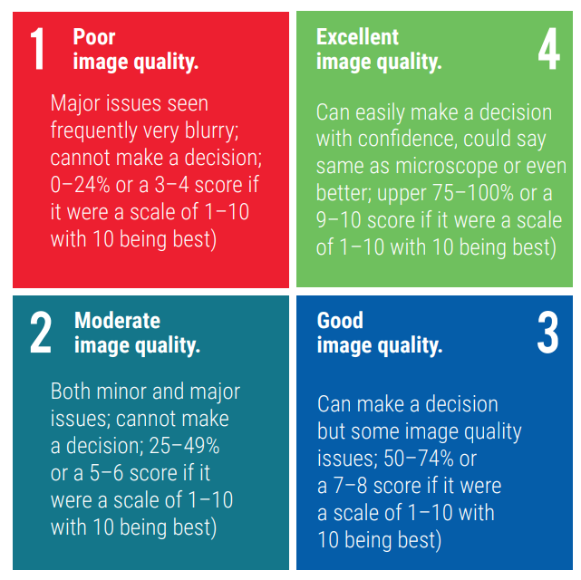
There was a minimum of a 2 week wash out period between the reads on images generated from Aperio AT2 and Aperio GT 450. Image quality was assessed by each participating Pathologist using a quadrant-based scoring criteria, as shown in Figure 1. For example, a score of 4 was recorded by the pathologist if he or she assessed that the image quality fit with the description in quadrant 4.
Results
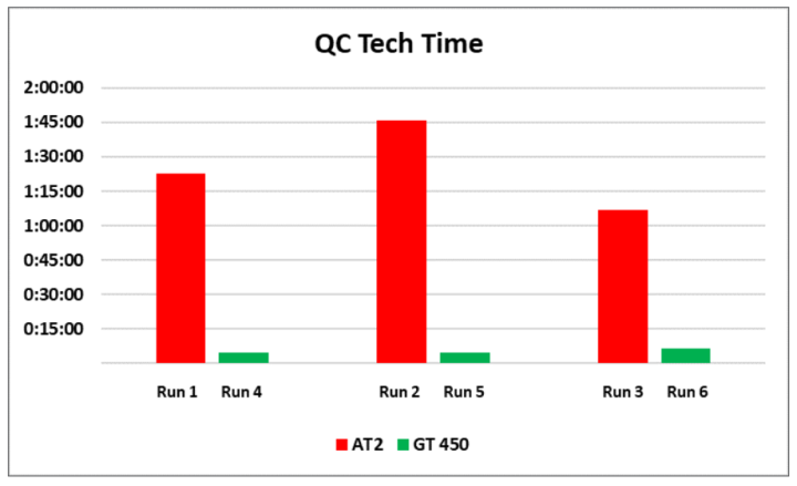
Total tech QC time
94% reduction.
QC tech time is defined as the aggregate time the operator spent with the instrument. This is the time that was required for slide quality or slide inspection purposes and is a subset of the total hands-on time.
Table 1 (below): Average Tech QC time was reduced from about 1 hour and 25 minutes to only 5 minutes with Aperio GT 450. This is a 94% reduction in Tech QC time.
| AT2 | Run 1 | Run 2 | Run 3 | Avg |
|---|---|---|---|---|
| Tech QC Time (h:mm:ss) | 1:22:38 | 1:45:41 | 1:06:46 | 1:25:02 |
| GT 450 | Run 4 | Run 5 | Run 6 | Avg |
|---|---|---|---|---|
| Tech QC Time (h:mm:ss) | 0:04:54 | 0:04:38 | 0:06:43 | 0:05:25 |
For Research Use Only. Not For Use In Diagnostic Procedures.
About the presenters
Lance Mikus earned his bachelor’s degree from UCLA. He worked as a scientist and lab manager at the UW-Madison Medical School, resulting in several publications, and went on to earn an MBA focused on Entrepreneurship and Marketing from the UW-Madison School of Business. Mr. Mikus has 20 years of commercial operations experience in the healthcare industry. He also holds a green belt in six sigma quality practices, is a certified practitioner in launch excellence and has been awarded 5 patents.
Ashley Troutman has been involved in Laboratory Medicine for more than 20 years in clinical, research and administrative capacities. He has worked in facilities of all sizes, from small community hospitals and private labs to large academic medical centers and corporate reference labs. He has extensive experience in laboratory science and management, specifically in anatomic pathology and immunohistochemistry. He has managed routine histology operations and has been part of the team to aid researchers in designing experiments using histologic techniques. These roles have allowed Ashley to lead work process implementation teams that saw success in scientific innovation as well as improving laboratory efficiency through areas of waste/cost reduction, process improvement and safety.
Julie Broccardo is the Director of Pathology Operations for NeoGenomics Laboratories, Inc. She is responsible for managing multi-site operations, including the Ft. Meyers, Houston, Carlsbad, and Aliso Viejo laboratories. Julie is passionate about developing her staff and working on initiatives to build and develop a more efficient lab.
Dr. Chizhevsky is the Senior Hematopathologist and Medical Director, Flow Cytometry, NeoGenomics Laboratories, Inc. He is a member of the College of American Pathologists, American Society for Clinical Pathology and International Clinical Cytometry Society. Dr.Chizhevsky has authored many publications in medical journals.
Related Content


Leica Biosystems content is subject to the Leica Biosystems website terms of use, available at: Legal Notice. The content, including webinars, training presentations and related materials is intended to provide general information regarding particular subjects of interest to health care professionals and is not intended to be, and should not be construed as, medical, regulatory or legal advice. The views and opinions expressed in any third-party content reflect the personal views and opinions of the speaker(s)/author(s) and do not necessarily represent or reflect the views or opinions of Leica Biosystems, its employees or agents. Any links contained in the content which provides access to third party resources or content is provided for convenience only.
For the use of any product, the applicable product documentation, including information guides, inserts and operation manuals should be consulted.
Copyright © 2026 Leica Biosystems division of Leica Microsystems, Inc. and its Leica Biosystems affiliates. All rights reserved. LEICA and the Leica Logo are registered trademarks of Leica Microsystems IR GmbH.