
High-Resolution Episcopic Microscopy (HREM) using the HistoCore NANOCUT R Rotary Microtome by Leica Biosystems
Abstract
High-resolution episcopic microscopy (HREM) is a 3D analytical technique based on volume data sets composed of consecutive digital images obtained from the block surface of methacrylate resin-embedded embryos or tissues, as they are physically sectioned. Since the images are captured from the face of the block of resin (in which the biological sample has been embedded) rather than from glass-mounted sections, the set is inherently complete, aligned and unfolded. HREM uses the fluorescence of eosin for providing morphology. Although the technique is based on episcopic images, these are of high resolution and close to histological quality: images reflect the tissue structure and densities revealed by histology, albeit in grey scale color map.
An HREM prototype was developed by the IGBMC imaging center and the ICS embryology/anatomopathology service (Illkirch, France) including the HistoCore NANOCUT R automated rotary microtome developed by Leica Biosystems. Blocks were tested for reproducibility of slice thickness, quality (especially of the block surface), and more importantly for the repositioning of the block in its 3D mode. We demonstrated that all the parameters tested fitted the objectives. Indeed, the regularity of sectioning was efficient, the surface of the block after each slice was highly smooth with no artifacts and finally the repositioning of the block was highly accurate permitting immediate 3D reconstruction of the samples without requiring a post-acquisition realignments step.
Introduction
Histological analyses of biological samples represent the gold standard in terms of resolution, image contrast and sensitivity to describe morphological defects. Although laborious and time-consuming, this technique is still applied for a lot of analysis requiring structural description. Over the past decades, there has been development of cutting-edge imaging tools to discover novel structural and functional information and imaging has revolutionized biomedical research.
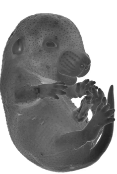
A major driver for the adoption of novel imaging techniques is the specific and varied requirements of individual research groups. For example, in the field of embryonic phenotyping, abnormal morphogenesis of complex developing structures in a developing embryo can be difficult to visualize by external examination or 2D sections of the embryo. Thus, the ability to record three-dimensional (3D) digital images of mutants at different developmental stages by using medium-to-high throughput technologies adds value to the information provided by histology. It allows automated detection of several defects by comparison with wild-type standards as well as the quantification of morphological changes (Adams et al., 2013). Besides the embryology field, examining organ shapes of adult models in 3D stacks can be advantageous because phenotypes can be easier to detect than in 2-dimensional histological sections, particularly for researchers with little anatomical training.
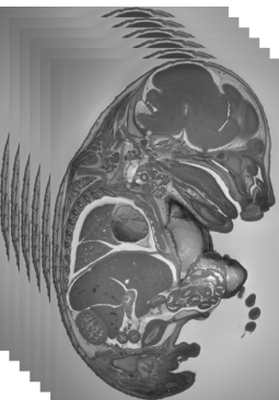
HREM is an effective and powerful tool for assessing structural abnormalities in embryos or organs ex vivo. Essentially ‘3D histology,’ a series of successive 2D images of the sample block face, is captured during histological sectioning of fixed and embedded tissues, providing structural information at high resolution with details approaching those conventionally obtained using histological analysis. HREM data can be analysed either as 2D image stacks or by 3D rendering, and they can reveal alterations in morphology that are impossible to detect using lower resolution techniques such as micro-CT or MRI. The voxel resolution of 3D models is only limited by the thickness of sections successively removed from the block surface. In practice, spatial resolutions of 1µm are standard (Weninger et al., 2006; Weninger and Mohun, 2002).
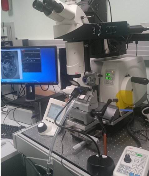
The accuracy of image alignment critically depends on the block face returning precisely to the same position for photography after each section cycle. This specification is not standard for rotary microtomes, which require modifications to ensure this capability. Some sledge microtomes were engineered to possess suitably precise stopping positions for photography (e.g., Leica SM2500), but they are no longer commercialized. Recently, Leica Biosystems developed a new rotary microtome HistoCore NANOCUT R that can be coupled with the optics and camera.
Experimental Procedures
Principle of the Procedure
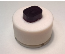
Phenotyping mouse embryos or organs using HREM requires the sample to be embedded in resin. Samples are gradually dehydrated in ethanol, before being embedded in JB4 resin to produce a block suitable for cutting. In order to produce contrast, the embedding resin is completed with Eosin B (emits fluorescence when mixed in JB4) and Acridine Orange (absorbs Eosin signal and creates contrast). Eosin staining is well known from histology and Acridine Orange has a strong affinity for nucleus, resulting in a contrast that strongly resemble to a grayscale capture of Hematoxylin & Eosin staining.
The embedded sample is placed on the HREM HistoCore NANOCUT R’s microtome by Leica Biosystems and lit with blue light (450-500 nm range) to acquire an image, then a slice is cut from the block to remove matter and the process is repeated. As the concentration of Eosin in the resin is very high, excitation light is absorbed within a micrometer from the block’s surface. This effect allows you to consider the images obtained from HREM as surface data (the data collected will be restricted to a depth below 1µm, and thus will never exceed the thickness of the section ≥ 1µm). The thickness of the sections will be adjusted to the pixel size of the images in order to obtain isotropic voxels (same size in x, y, z dimensions).
The result is a series of images taken across the sample that can be used to reconstruct image data in 3D, allowing you to examine a slice in any orientation within the stack without loss of quality.
Please note that HREM processing is destructive, the sample being cut into sections, and that one sample can therefore only be imaged once. This makes the initial setup of the sample critical to get optimal images that will serve sample analysis the best.
Materials
- Biological material: Mouse embryos or fetuses, pieces of adult tissues no larger than 20 mm.
- Bouin’s fixative
- JB-4 resin embedding kit (Polysciences, Inc.)
- Eosin B (Sigma-Aldrich)
- Acridine Orange (Sigma-Aldrich)
- Ethanol 100% and a series of 70%, 80%, 90%, 95% in H2O
- Polyethylene embedding molds
- TC-65 Tungsten Carbide blades (Leica Biosystems)
Sample Embedding
This protocol is simplified to illustrate the basics of the procedure. For detailed protocol see https://dmdd.org.uk/hrem/sample-preparation/
- Fix the specimens in Bouin’s fixative
- Wash in graded ethanol solutions: 70%, 80%, 90%, 95%, 100%
- Prepare JB4 resin mixture containing Acridine orange and eosin.
- Incubate in mixed resin/ethanol (1:1)
- Incubate in pure resin mixture
- Prepare fresh activated JB4 resin
- Polymerize a cushion in the bottom of the mould
- Place the sample on the cushion in the mould containing activated resin and polymerize the resin at 4°C overnight. The resin is covered with mineral oil to avoid contact with the air.
- Further harden the block in an oven at 95°C
Locate the sample within the block using an oblique illumination and trim the resin that does not contain the specimen (will permit to adjust at the better magnification)
Sectioning Procedure
- Mount the block on the HistoCore NANOCUT R microtome by Leica Biosystems
- Use 3D mode of the microtome
- Trim block until the very beginning of the specimen
- Adjust the focus
- Start the automated sectioning and image capture. Special adapted software modules (MetaMorph® Software or home-made software available upon request) will permit the synchronization of cutting and image capturing.
- Sections can be performed from 2µm to 7µm. As an example, embryos at E15.5 stage are cut at 7µm.
- For data processing, optimize grayscale mapping as desired; scale data and/or subsample as appropriate for 3D rendering software.
Results
A. Prototype Apparatus for Episcopic Imaging
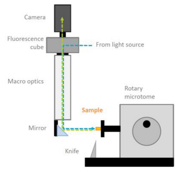
The HREM prototype is composed of a camera SCMOS, a GFP- fluorescence filter, a LED light source, optical device (macroscope), a mirror, the knife holder and the sample mounted on the HistoCore NANOCUT Rotary microtome by Leica Biosystems.
B. Quality and Regularity of Sectioning
Even if only the images are captured from the face of the block of resin, the sections are physically done meaning that cutting artefacts can be visible on the pictures. The resin blocks are sectioned in steps from 2µm to 7µm. TC-65 tungsten carbide blades (Leica Biosystems) were used to section the blocks. On the three blocks that were cut with the HistoCore NANOCUT R by Leica Biosystems, the surface of the block was perfect with no artefact observed (see figure 3).
C. 3D reconstruction
The novelty of the HistoCore NANOCUT R rotary microtome by Leica Biosystems resides in its 3D optional mode. This means that compared to other microtomes, it was modified to allow the block holder to reproducibly come to rest at a “photo-position” after each cut. The more accurate the position is obtained, the more accurate the alignments are obtained and the best 3D reconstruction obtained. HREM data can be analysed either as 2D image stacks or by 3D rendering using appropriate software. Thanks to alignment of the images and achievement of isotropic pixels, orthogonal and oblique views of the tissue sample can be obtained from HREM data. With the blocks that were tested, the “photo-position” was perfectly obtained using the 3D mode of the HistoCore NANOCUT R microtome. The 3D reconstruction was so good that realignment taking a step after acquisition was not necessary.
Discussion
The development of the HistoCore NANOCUT R microtome by Leica Biosystems with a 3D option is of great interest to people who wish to develop HREM technology in their laboratory. Performing automated sections using the Leica HistoCore NANOCUT R microtome led to increased reproducibility of key parameters like the cutting surface and precise positioning of the block leading to perfect alignment of the sections and enhanced quality of 3D reconstructions.
HREM digital images offer several advantages compared to conventional histological sections. Firstly, they do not show distortions introduced by the sectioning, section processing or mounting processes. Furthermore, a series of thousands of images are produced within a few hours in an automated way in contrast to weeks to produce a series of more or less precisely realigned digital images from a thousands of histological sections. HREM volume data can be immediately analysed with orthogonal and oblique virtual re-sectioning tools and appropriate software.
HREM has proved to be an excellent tool in a broad variety of research fields. In addition to the increasing use of HREM for visualizing embryos for screening the phenotype of prenatally lethal E14.5/E15.5 mouse embryos produced for the International Mouse Phenotyping Consortium (IMPC) (Dickinson at al., 2016),it was very recently also employed for visualizing tissue samples of adult biomedical model organisms (Weninger et al., 2013; Geyer et al., 2015), human skin biopsies (Tinhofer et al., 2018) and biological materials used in modern medicine. The highly encouraging results lead us to the conclusion that HREM has the potential to become a valuable tool in various fields of pre- and clinical research within the next decade.
Therefore, HREM offers low-cost and high-quality information using readily available laboratory equipment, which is particularly important for individual researchers.
Acknowledgements
We thank Dr T. Mohun and W.J. Weninger for a constructive discussion and their help in the initial step of this project. The project was supported by the French state funds through the “Agence Nationale de la Recherche” under the frame programme “Investissements d’Avenir” labelled (ANR-10-IDEX-0002-02, ANR-10-LABX-0030-INRT, ANR-10-INBS-07 PHENOMIN).
The following research was prepared by an independent third-party on behalf of Leica Biosystems. Leica Biosystems was not involved in preparing this research, which reflects an independent, outside opinion.
Olivia Wendling1-5, Didier Hentsch1-4, Fabien Pertuy1-5, Hugues Jacobs1-5, Nicolas Lemercier 1-4, Serge Taubert1- 4 Jean-Luc Vonesch1-4, Patrick Reilly1-5, Bertrand Séraphin1-4, and Yann Hérault1-5
- Institut de Génétique et de Biologie Moléculaire et Cellulaire, Illkirch, France
- Center National de la Recherche Scientifique, UMR7104, Illkirch, France
- Institut National de la Santé et de la Recherche Médicale, U1258, Illkirch, France
- Université de Strasbourg, Illkirch, France
- ICS Infrastructure, Illkirch, France
References
Adams D, Baldock R, Bhattacharya S, Copp AJ, Dickinson M, Greene ND, Henkelman M, Justice M, Mohun T, Murray SA, Pauws E, Raess M, Rossant J, Weaver T, West D. Bloomsbury report on mouse embryo phenotyping: recommendations from the IMPC workshop on embryonic lethal screening. Dis Model Mech. 2013 May;6(3):571-9.
Dickinson, M.E., Flenniken, A.M., Ji, X., Teboul, L., Wong, M.D., White, J.K., Meehan, T.F., Weninger, W.J., Westerberg, H., Adissu, H., et al. (2016). High-throughput discovery of novel developmental phenotypes. Nature 537, 508-514.
Geyer SH, Tinhofer IE, Lumenta DB, Kamolz LP, Branski L, Finnerty CC, Herndon DN, Weninger WJ. High-resolution episcopic microscopy (HREM): a useful technique for research in wound care. Ann Anat. 2015 Jan.
Mohun TJ, Weninger WJ. Embedding embryos for high-resolution episcopic microscopy (HREM). Cold Spring Harb Protoc. 2012 Jun 1;2012(6):678-80.
Mohun TJ, Weninger WJ. Generation of volume data by episcopic three-dimensional imaging of embryos. Cold Spring Harb Protoc. 2012 Jun 1;2012(6):681-2.
Mohun TJ, Weninger WJ. Episcopic three-dimensional imaging of embryos. ColdSpring Harb Protoc. 2012 Jun 1;2012(6):641-6.
Tinhofer IE., Zaussinger M., Stefan H. Geyer SH., Meng S., Kamolz LP., Tzou CHJ. and Weninger WJ. The dermal arteries in the cutaneous angiosome of the descending genicular artery. Journal of Anatomy. 2018
Weninger WJ, Mohun T. Phenotyping transgenic embryos: a rapid 3-D screening method based on episcopic fluorescence image capturing. Nat Genet. 2002 Jan;30(1):59-65.
Weninger, W. J., et al. High-resolution episcopic microscopy: a rapid technique for high detailed 3D analysis of gene activity in the context of tissue architecture and morphology. Anat Embryol. 211 (3), 213-221 (2006).
Weninger WJ., Kamolz LP and Geyer SH. 3D visualization of Skin Substitutes. In: Kamolz L. P., Lumenta D. B., editors. Dermal Replacements in General, Burn, and Plastic Surgery. Springer, Wien (2013).
Related Content


Leica Biosystems content is subject to the Leica Biosystems website terms of use, available at: Legal Notice. The content, including webinars, training presentations and related materials is intended to provide general information regarding particular subjects of interest to health care professionals and is not intended to be, and should not be construed as, medical, regulatory or legal advice. The views and opinions expressed in any third-party content reflect the personal views and opinions of the speaker(s)/author(s) and do not necessarily represent or reflect the views or opinions of Leica Biosystems, its employees or agents. Any links contained in the content which provides access to third party resources or content is provided for convenience only.
For the use of any product, the applicable product documentation, including information guides, inserts and operation manuals should be consulted.
Copyright © 2026 Leica Biosystems division of Leica Microsystems, Inc. and its Leica Biosystems affiliates. All rights reserved. LEICA and the Leica Logo are registered trademarks of Leica Microsystems IR GmbH.