63 result(s) for 'Fluorescent'
41 - 50 of 63 results for 'Fluorescent'
Sort by
Show

The content of this webinar will encompass detailed technical descriptions of novel and state of the art methods to decalcify tissue samples that will be embedded in paraffin or for frozen sectioning for molecular and nucleic acid analysis for basic research...

In this session, we will briefly review the basics of molecular biology, examine critical factors which affect the quality of nucleic acids in the tissues and cells which are submitted for downstream molecular diagnostics, and briefly introduce some...

Different types of cancers frequently metastase to bone tissue. Treatment planning decisions are often based upon histology and special staining of these distant sites of disease. These decisions may rely on the outcome of immunohistochemistry, in situ...

The potential for non-invasive tests that provide equivalent research and diagnostic value as can be obtained from tissue biopsies is real, but not yet realized. Tissue biopsies allow for identification, phenotyping and molecular analysis of cancer and...
Cytogenetics has played a pivotal role in clinical and cancer diagnostics for over 60 years. The use of genomic technologies speeds targeted genetic analysis - but can high throughput automation in the cytogenetics laboratory broaden traditional approaches?

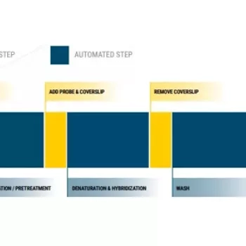
When was the last time that your pathologist brought you a slide of decalcified bone, and said it was the best she ever saw? Ever wonder why your PAS stain is not staining the basement membrane the way it should? These questions and 18 others will be discussed...

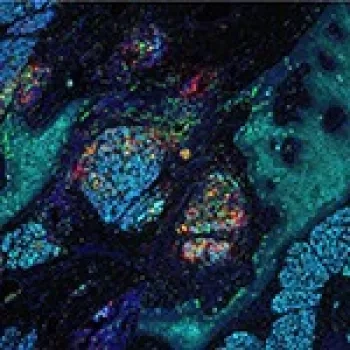

Dean joined Leica Biosystems in 2016 and is a senior scientist in the R&D team in Melbourne, working on advanced staining projects. He graduated from Monash University where he pursued a PhD, with the department of Immunology and Pathology. During this time, he investigated the role of T cell lineages in mediating inflammatory retinal disorders. He assessed cellular interactions using fluorescent multiplex IHC with confocal microscopy imaging of retinal wholemounts.