
Buying a Research Microtome: Why Quality Matters in Research Microtomy
Quality matters. In the research field, data quality is king. High-quality, reproducible, and reportable results matter when racing against manuscript publication and grant funding deadlines. Primary Investigators and researchers performing tissue-based research depend on the laboratory's workflow excellence, providing well-processed sections for data development.
What happens when murky data results from variability in microtomy techniques?
Improper instrument selection or configuration and an unclear understanding of what type of sectioning the lab will be doing can lead to delayed results or poor-quality data. Delays such as these can cause a ripple effect for investigators, threatening result credibility. Leica Biosystems can help optimize your sectioning workflow, allowing users of all experience levels to choose their sample parameters and enhancing sample adequacy.
Automating innovation: Delivering today's tissue-based research-driving tomorrow's clinical outcomes
Important Considerations for Research Microtomy
It is important to consider two key factors when preparing your research laboratory to perform microtomy. The first key consideration is determining what sample sources your research requires is critical to choosing the right microtome for your applications. The second key consideration is defining and using the proper microtome configurations to ensure downstream sample adequacy.
Choosing the microtome that lets you get the best possible section from every block and broadens your research with a wide selection of blade and specimen holders enables you to discover breakthroughs in research for biomedical to industrial applications.
There are three challenges researchers face when adding microtomy into their research laboratory:
- Identifying the application type and kind of materials used for the research project
- Understanding what type of microtome is needed for their research
- Knowing how to configure the microtome for optimal use properly
Considerations for choosing a microtome
Applications
Knowing what type of application your research requires is a first step to understanding what type of microtome will be the best fit for your laboratory.
First, define the purpose of the experiments and the type of specimen to be cut in your research plan. What goal or problem do you wish to solve by cutting a material?
Second, determine the density and thickness of the materials to be sectioned; will they be soft, like those of tissue in paraffin, hard, like bone or teeth in resin, or plastic materials used in manufacturing?
Third, having clear parameters on the purpose of the experiment, specimen, and type of material you plan to cut will help determine the correct knives, blades, and angle to use; options include Fixed or disposable metal, glass, and diamond blades.
Manual, semi-automatic, or fully automatic rotary microtomes?
Each element of choosing the type of microtome needed in your laboratory has its merits.
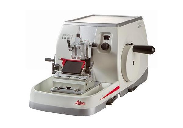
A manual microtome uses a specimen feed and cutting stroke (or rotary motion of the wheel) that is managed by hand. It also has a lower throughput, but this is not always detrimental. The ability to manually cut and evaluate each section offers microtomists the opportunity to concentrate on each block and section with greater control over the cutting process. The ability to assess in real time provides potential time savings through consistency. Still, this option requires a more significant amount of vigilance on behalf of the user to maintain quality standards.
A semi-automated microtome uses a specimen feed that is motorized, but the rotary motion of the handwheel is still done manually and offers a medium throughput. The semi-automated method works well for thick and hard sections, using a consistent force when cutting through a block. Automating the specimen feed provides the precision of a motorized specimen feeding that yields consistent, reproducible sections.
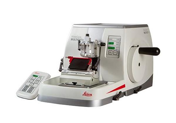
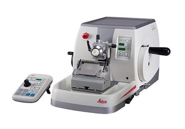
A fully automated microtome has both specimen feed and rotary motion of the handwheel automated, contributing to the ability to produce high-throughput sectioning. The mechanization of the instrument allows for automated setting and correction of feed movement and knife cutting. In addition, automation also provides consistency from user to user, so it can speed up time and provide reproducibility of sectioning. Many fully automatic microtomes allow for cutting a more comprehensive range of thicknesses, providing researchers flexibility when choosing sectioning materials such as paraffin or resin-based fillers.
Guides for microtome selection and configurations
Leica Biosystems is your trusted workflow partner, helping you obtain the best research data on various microtomy applications. Below are two helpful tools, a microtome buyer's guide, and a configuration guide. These guides are intended to help you select and configure the right microtomes for your application types, including human and non-human specimens embedded in paraffin, undecalcified bone, and automotive and plastic parts.
How do I know what is right for my laboratory?
Let us guide you in selecting the right features based on your research applications.
The Leica Biosystems Research Microtome Comparison Guide provides help in selecting the right features based on your research applications. Together with the original validated accessories, our reliable and precise microtomes are designed to provide high-quality sections and enhanced efficiency while maintaining a safe and healthy workplace.
What are the important features when choosing a microtome?
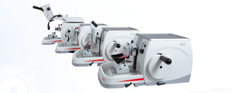
This easy-to-use microtome can help guide to help you through the decision-making process. Leica Biosystems rotary microtomes offer microtomists the precision, control, and comfort they need to get the best from every block. Our rotary microtomes provide the consistency required to deliver high-quality sectioning and maintain optimized workflows.
-
Image
-
Image
-
Image
-
Image
| TECHNICAL DATA | ||||
| Type of microtome | Mechanical | Semi Motorized | Fully Motorized | Fully Motorized |
| GENERAL | ||||
| Nominal supply voltages: | N/A | 100 / 120 / 230 / 240 V AC | 100 / 120 / 230 / 240 V AC | 100 / 120 / 230 / 240 V AC |
| Nominal frequency : | N/A | 50/60 Hz | 50/60 Hz | 50/60 Hz |
| DIMENSIONS AND WEIGHT | ||||
| Width (including handwheel and coarse feed wheel) Depth (including the section waste tray) Height (without top tray) W x D x H: |
477 mm x 620 mm x 295 mm | 477 mm x 620 mm x 295 mm | 477 mm x 620 mm x 295 mm | 415 mm x 620 mm x 295 mm |
| Weight (without accessories): | Approx. 31 kg | Approx. 31 kg | Approx. 40 kg | Approx .40 kg |
| MICROTOME | ||||
| Section thickness setting range: | 1 - 60 µm | 0.5- 100 µm | 0.5 - 100 µm | 0.25 - 50 µm |
| Trimming section thickness setting range: | 10 µm, 30 µm | 1 - 600 µm | 1 - 600 µm | 1 - 300 µm |
| Specimen feed: | Approx. 24 mm ±2 mm | Approx. 24 mm ±1 mm | Approx. 24 mm ±1 mm | Approx. 24 mm ±1 mm |
| Vertical stroke: | 70 mm ±1 mm | 70 mm ±1 mm | 70 mm ±1 mm | 70 mm ±1 mm |
| Maximum specimen size (H x W x D): |
Large standard clamp: 55 x 50 x 30 mm Super Cassette clamp: 68 x 48 x 15mm |
Large standard clamp: 55 x 50 x 30 mm Super Cassette clamp: 68 x 48 x 15 mm |
Large standard clamp: 55 x 50 x 30 mm Super Cassette clamp: 68 x 48 x 15 mm |
Large Standard clamp: 55 x 50 x 30 mm Super Cassette clamp: 68 x 48 x 15 mm |
| Unique force balance system | Yes | Yes | Yes | Yes |
| Specimen retraction: | Approx. 40 µm; can be turned off | 5 - 100 µm in 5 µm increments; can be turned off | 5 - 100 µm in 5 µm increments; can be turned off | 5 - 50 µm (in 5 µm increments); can be turned off |
| COARSE FEED AND MOTORIZED SECTIONING SPEEDS | ||||
| Slow forward and backward speed Fast-forward speed Fast backward speed (fast homing) |
N/A | 300 µm/s 800µm/s 1800 µm/s |
300 µm/s 800µm/s 1800 µm/s |
150 µm/s 400µm/s 900 µm/s |
| Sectioning speed: | N/A (manual) | N/A (manual) | O - 420 mm/s ±10 % | o - 195 mm/s ±10% |
| Personalized coarse feed wheel | User selectable | User selectable | User selectable | NIA |
| Specimen orientation with zero position horizontal / vertical rotation: | ± 8°/± 8° | ± 8°/± 8° | ± 8°/± 8° | ± 8°/± 0° |
| Waste tray | Standard | Standard | Standard | Standard |
For Research Use Only. Not for use in diagnostic procedures.
How do I know what the best configuration for my research application is?
Our Microtome Configuration Guide will help you choose the right microtome for the job.
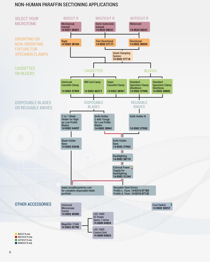
Does your laboratory's research require the sectioning of non-human biological specimens embedded in paraffin, undecalcified bone, automotive or plastic parts?
Our reliable and precise microtomes, in combination with the original validated accessories, are designed to provide high-quality sections and enhanced efficiency while maintaining a safe and healthy workplace.
Choose the rotary microtome that lets you get the best possible section from every block and broaden your research with a wide selection of blade and specimen holders, enabling you to discover new breakthroughs in research for biomedical to industrial applications.
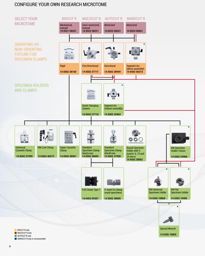
Leica Biosystems can help your custom configure your microtome requirements based on your research needs in a few easy steps. Answer a few questions, then follow the schematic to help choose the correct application:
Step 1: Configure your research microtome type:
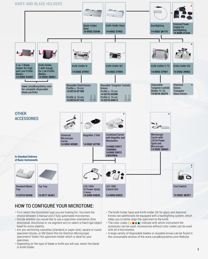
Step 2: Select your knife and blade holders:
Step 3: Non-Human sectioning applications parameters:
Conclusion
Together with the original validated accessories, our reliable and precise microtomes are designed to provide high-quality sections and enhanced efficiency while maintaining a safe and healthy workplace.
Having a 145+ year heritage in tissue workflow diagnostics, Leica Biosystems is uniquely suited to fit research laboratories with customized solutions that continuously ensure high-quality, reproducible, and reportable data - from research to result.
Related Content


Die Inhalte des Knowledge Pathway von Leica Biosystems unterliegen den Nutzungsbedingungen der Website von Leica Biosystems, die hier eingesehen werden können: Rechtlicher Hinweis. Der Inhalt, einschließlich der Webinare, Schulungspräsentationen und ähnlicher Materialien, soll allgemeine Informationen zu bestimmten Themen liefern, die für medizinische Fachkräfte von Interesse sind. Er soll explizit nicht der medizinischen, behördlichen oder rechtlichen Beratung dienen und kann diese auch nicht ersetzen. Die Ansichten und Meinungen, die in Inhalten Dritter zum Ausdruck gebracht werden, spiegeln die persönlichen Auffassungen der Sprecher/Autoren wider und decken sich nicht notwendigerweise mit denen von Leica Biosystems, seinen Mitarbeitern oder Vertretern. Jegliche in den Inhalten enthaltene Links, die auf Quellen oder Inhalte Dritter verweisen, werden lediglich aus Gründen Ihrer Annehmlichkeit zur Verfügung gestellt.
Vor dem Gebrauch sollten die Produktinformationen, Beilagen und Bedienungsanleitungen der jeweiligen Medikamente und Geräte konsultiert werden.
Copyright © 2024 Leica Biosystems division of Leica Microsystems, Inc. and its Leica Biosystems affiliates. All rights reserved. LEICA and the Leica Logo are registered trademarks of Leica Microsystems IR GmbH.