Lung PD-L1 AI
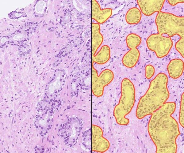
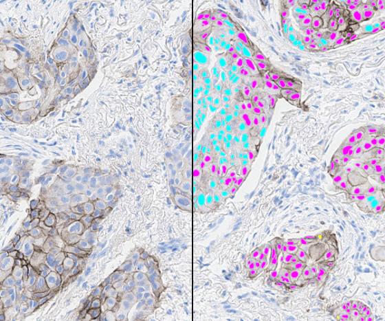
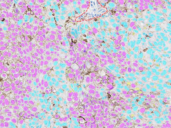
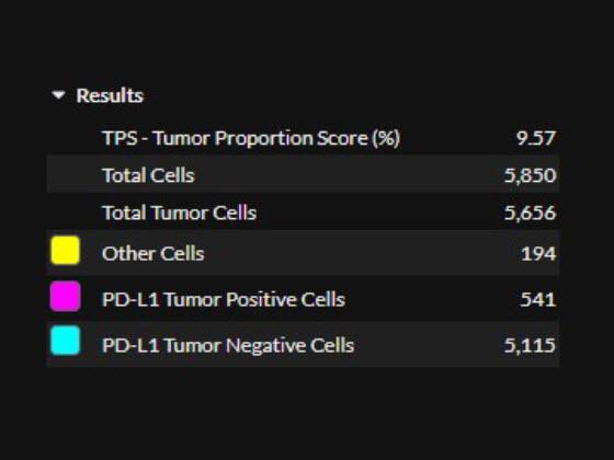
Developed by Indica Labs, the Lung PD-L1 AI solution leverages deep learning that standardizes scoring of PD-L1 IHC in non-small cell lung cancer (NSCLC) tissue samples. The algorithm reports quantitative results and markup images, including tumor detection, cell phenotyping based off the PD-L1 staining, and tumor proportion score (TPS).
Lung PD-L1 AI is For Research Use Only and not intended for clinical diagnostic use. Lung PD-L1 AI accessed via the Aperio HALO AP image management system.

Lung PD-L1 AI in Aperio HALO AP Demonstration
Explore the future of pathology with the Lung PD-L1 AI app from Indica Labs. Lung PD-L1 AI seamlessly integrates within the Aperio HALO AP enterprise digital pathology platform to identify PD-L1 positive and PD-L1 negative tumor cells in NSCLC tissue.
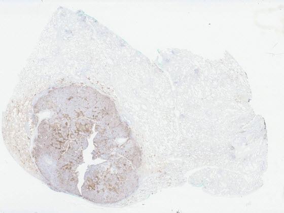
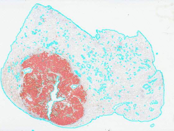
After selecting an image, view the IHC slide in the viewer and the Lung PD-L1 AI results. Results include overlays and quantitative results. The first set of overlays prepare the tissue for analysis by identifying areas of analyzable tissue. The next overlay identifies areas of tumor in the sample. The final overlay identifies PD-L1 positive and negative tumor cells within the tumor area. Finally, quantitative results are displayed in the assay panel to the right and include a tumor proportion score (TPS).
