
Assessing Timing and Efficiency: A Comparative Evaluation of the Aperio GT 450 DX Scanner and a High-throughput Digital Slide Scanner
Digital Pathology can offer agility to Anatomic Pathology departments who are invested in improving efficiencies and high levels of quality while quickly responding to the increasing pressure of on-demand pathology services. While there are many factors that can affect the overall impact of digital pathology, in this study, we are focusing on instrument operation. Our goal is to compare and quantify the precise amount of automated time required to digitise slides between the Aperio GT 450 DX and a commonly used, high-throughput digital slide scanner.
The Aperio GT 450 DX is an automated, high-capacity digital pathology slide scanner made by Leica Biosystems. The Aperio GT 450 DX generates an output of 81 slides/hour at 40x, delivering up to 450 scanned slides in one batch.
The high-throughput digital slide scanner used for comparison generates an output of more than 82 slides/ hour at 40x, delivering up to 360 scanned slides in one batch.
Method
This study, conducted by the Leica Biosystems Content and Evidence Team, occurred at a University Medical Centre in Europe. This site processes approximately 300,000 slides annually and was chosen due to experience in creating and using digitised images to perform primary diagnosis.
A Lean and Six Sigma time and motion analysis was performed to evaluate the time required to digitise a full set of glass slides. A set of 30 slides, representative of the site’s daily work, was selected by the laboratory staff to be scanned on both digital scanners. Hands-on time and instrument operation time were captured across the entirety of the process: including loading the slides into each respective rack, slide digitisation, and unloading the slides from the scanner. After scanning was complete, if required, the laboratory staff verified the quality of each digitised slide image. Timing data was pulled from each scanning instrument’s log file and compared to the time and motion benchmark data to ensure alignment and accuracy.
As the laboratory team participating in the study regularly processes multiple tissue types, the glass slides scanned included a wide variety of tissue, including: stomach, cervix, bone marrow, skin, and kidney biopsies plus liver, thyroid, and lung resections. Additionally, multiple staining techniques were utilised, including: haematoxylin and eosin (H&E), Grocott’s Methenamine Silver (GMS), Giemsa, Periodic Acid-Schiff (PAS), Alcian Blue, and several immunohistochemical stains (ER, PR, CD10, SOX-10).
Results
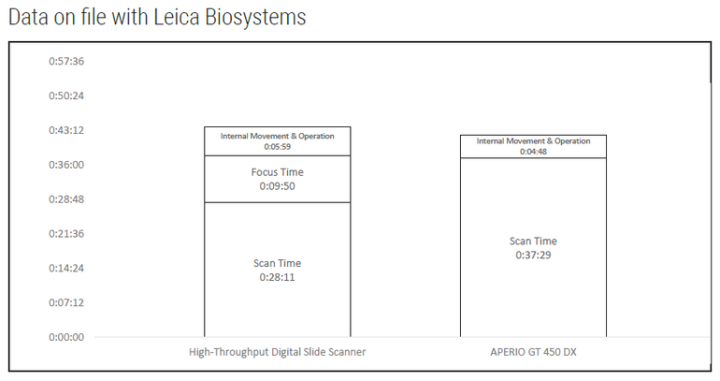
The data from each scanning instrument is summarised in the following figures.

Discussion
Upon initial review of scan times, the commonly used, high-throughput digital slide scanner appears to be much faster at 28:11 (mm:ss) than the Aperio GT 450 DX at 37:29 (mm:ss). Upon further investigation, the time and motion study revealed that the high-throughput digital slide scanner must perform two separate operations to generate a digitised image. These operations included scanning, for 28:11 (mm:ss), and focusing, for 9:50 (mm:ss), resulting in a total time of 38:01 (mm:ss). A review of the high-throughput digital slide scanner log file validated this finding. In contrast, the Aperio GT 450 DX performs scanning and focusing operations simultaneously for a total time of 37:29 (mm:ss).
Furthermore, we found that each scanning platform requires additional internal movements and processing steps, adding 5:59 (mm:ss) processing time for the high-throughput digital slide scanner and 4:48 (mm:ss) processing time for the Aperio GT 450 DX.
Looking at the comprehensive automated slide digitisation process, the Aperio GT 450 DX was faster than the high-throughput digital slide scanner by 01:43 (mm:ss) for this common batch of 30 slides. For an Anatomic Pathology Laboratory with a comparable volume to this University Medical Centre, potential realisation of time savings could scale to 253 hours annually through use of the Aperio GT 450 DX.
Conclusion
Although scan speed is a key performance indicator used for digital scanning instruments, operators should be conscious that the reported scan speed is only one factor impacting overall effectiveness. Notably, scan times are often defined differently across digital scanning instruments and may have a downstream impact on realised efficiency. When comparing digital scanning instruments, it is important to consider all automated processing steps and times required to digitise slides, from slide loading to unloading
*Scan speed assumes 15mm x 15mm area at 40x.
** Scan speed assumes 15mm x15mm area at 20x and 40x, for the case of 5 focus points.
Projections and Realised Results are specific to the institution where they were obtained and may not reflect the results achievable at other institutions.
For In Vitro Diagnostic Use. The clinical use claims described for the products in the information supplied have not been cleared or approved by the U.S. FDA or are not available in the United States.
This reference document is presented as a service to health care professionals by Leica Biosystems and has been compiled from available literature. Although every effort has been made to report faithfully the information, Leica Biosystems cannot be held responsible for the correctness. This document is not intended to be, and should not be construed as medical advice. For any use, the product information guides, inserts and operation manuals of the various drugs and devices should be consulted. Leica Biosystems and the editors disclaim any liability arising directly or indirectly from the use of drugs, devices, techniques or procedures described in this reference document.
Projections and Realized Results are specific to the institution where they were obtained and may not reflect the results achievable at other institutions.
Want to see how the Aperio GT 450 Dx scanner can enhance your lab's efficiency?
Related Content

Leica Biosystems content is subject to the Leica Biosystems website terms of use, available at: Legal Notice. The content, including webinars, training presentations and related materials is intended to provide general information regarding particular subjects of interest to health care professionals and is not intended to be, and should not be construed as, medical, regulatory or legal advice. The views and opinions expressed in any third-party content reflect the personal views and opinions of the speaker(s)/author(s) and do not necessarily represent or reflect the views or opinions of Leica Biosystems, its employees or agents. Any links contained in the content which provides access to third party resources or content is provided for convenience only.
For the use of any product, the applicable product documentation, including information guides, inserts and operation manuals should be consulted.
Copyright © 2026 Leica Biosystems division of Leica Microsystems, Inc. and its Leica Biosystems affiliates. All rights reserved. LEICA and the Leica Logo are registered trademarks of Leica Microsystems IR GmbH.