The Power of Digital Pathology in Research

Learning Objectives
- The power of digital pathology and its benefits to the AP workflow
- The future of digital pathology
Webinar Transcription
The Power of Digital Pathology in Research
1. Leica Biosystems Proprietary Information. For Research Use Only. Not For Use in Diagnostic
The Power of Digital Pathology In Research. Caitríona Lyons PhD.
2. Introduction
- Caitríona Lyons, PhD
- Based in Dublin, Ireland
- Current Role: Product Manager Image Analysis, Leica Biosystems
- Previous Roles:
- Product Owner Image Analysis, Leica Biosystems
- Image Analysis Specialist, Leica Biosystems
- Education:
- BSc (Hons) Biomedical Science
- PhD Medicine and Health
- Postdoc Immunology
3. Agenda
- History of Pathology
- What is Digital Pathology
- Power of Digital Pathology
- Leica Biosystems Role in Digital Pathology
- The Future of Digital Pathology
- Final Thoughts
4. History of Digital Pathology
- Pathology has been around for over 100 years
- Telepathology has been around for nearly 50 years
- Past decade: a true digital transformation has been happening
- Whole slide imaging technology
- Advances in software applications
- Integration into different environments
5. What is Digital Pathology
- Acquisition, management, sharing and interpretation of pathology information in a digital environment
- High-resolution digital images are captured with a scanning device that can be viewed on a computer screen or mobile device
- Both bright-field and fluorescent images can be captured
6. Access to the right expertise on the right slide with the right tools
- Efficient workflows
- Remote access
- Ability to view your images and data from anywhere
- Freedom from the microscope
- Quantitative Image Analysis at a level of detail impossible by eye
- Maintain integrity of data
- Searchable digital archive
7. Tools to flag and help review complex or unusual slides / cases
- Annotation of slides
- Automatic micron measurement
- Easy, accurate measurement of features
- Number, color and intensity
- View multiple slides side-by-side
8. Maintains the integrity of the tissue on the slide
- High to low throughput of glass slides
- Walkaway scan protocol
- Scans almost any brightfield or fluorescent stains
- Supports 1x3 and 2x3 glass slide formats
9. Images and data generated from the scanner can be meaningfully organized into specimens, cases and projects
- Library of images generated from scanner is now:
- Searchable
- Accessible
- Controllable
10. Utilizes the managed images with image analysis applications to:
- Identify cohorts of cells
- Identify and score specific assays
- Phenotype cells
- Provides a permanent record of your results for every cell on a digital slide
- Results that are Reproducible
11. Identification of cohorts of cells, like infiltrating lymphocytes and tumor in breast H&E tissue
- Identification and Scoring of specific assays, such as PD- L1 membrane stain in annotated regions of interest of lung tumor
- Cell phenotyping, in this instance of cultured rat neurons based on antibody
12. MANAGE Deployment Options
- Enterprise Sharing and Expert Collaboration Aperio eSlide Manager (On-premise) Scanning & Hosting
- LOCAL Single site
- HUB & SPOKE Multi-site
- ANALYZE
- IHC & FL
- IHC
- Protein Biomarkers RNA ISH & FISH
- Molecular Biomarkers GENIE BF & FL Tissue Morphology
- SCAN OIL / FLUORESCENCE / BRIGHTFIELD
- Aperio CS5
- Aperio FL Scanning Systems
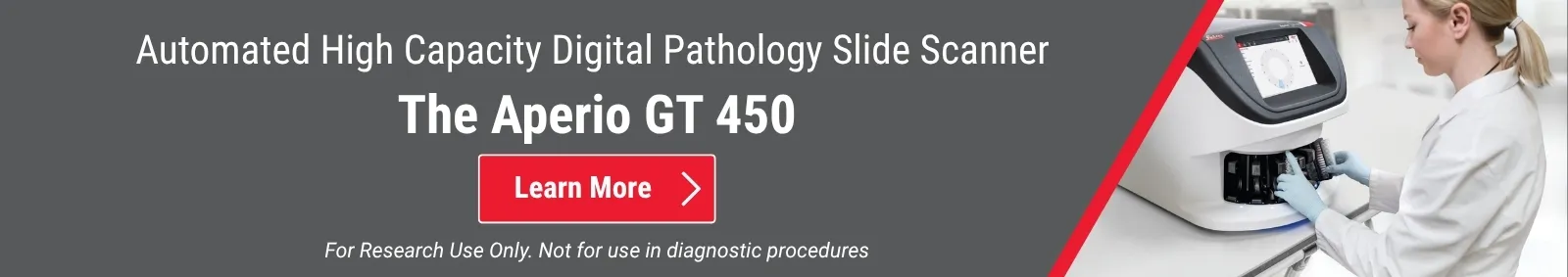
- Leica Biosystem’s Role in Digital Pathology Aperio GT 450 (High Throughput)
13. Uses Machine Learning
- Identification / Quantification of Disease vs Normal
- Uses include oncology, toxicology, nephrology, injury & inflammation
- Rapid / Consistent Annotation of Tissue(s)
- Heterogeneous samples
- Limited access to expert review
- Validation or Aperio iQC
14. Artificial Intelligence – AI John McCarthy in the 1955 proposal for the 1956 Dartmouth Conference “…every aspect of learning or any other feature of intelligence can in principle be so precisely described that a machine can be made to simulate it…” Machine Learning – ML Arthur Samuel, an IBM scientist, in 1959 “…field of study that gives computers the ability to learn without being explicitly programmed...” Deep Learning – DL 1965 – 1st deep learning model was developed by Alexey Grigoryevich Ivakhnenko and Valentin Grigorevich Lapa The Future of Digital Pathology: Artificial Intelligence
15. AI may provide the solution:
- Aids in research
- Automates assay(s)
- Automates Image Analysis
- Data is ready and available before the slide is open
Range of H&E supported H&E Tumor mask from Prototype Cytokeratin – additional ground truth to exploit
16. Removes bias by allowing quantitative analysis Allows you to access your images from anywhere Consolidates your physical slides with digital images Provides a permanent archive of images The Future is Bright, The Future is Digital
Leica Biosystems Proprietary Information. For Research Use Only. Not For Use in Diagnostic
Copyright © 2026 Leica Biosystems Imaging, Inc. All Rights Reserved. LEICA and the Leica logo are registered trademarks of Leica Microsystems IR GmbH. Aperio is a registered trademark of Leica Biosystems Imaging, Inc. in the USA and optionally in other countries. GT and GT 450 are trademarks of Leica Biosystems Imaging, Inc. in the USA and optionally in other countries. Other logos, product and/or company names might be trademarks of their respective owners.
200608 Rev. A IMC-8281-REV A
About the presenter
Caitriona earned a BSc in Biomedical Science and a PhD in Medicine and Health from University College Cork, Ireland. Her PhD focus was on Immunology, she studied potential cross-talk between apoptotic signalling pathways and innate immune signalling pathways.
Related Content


Leica Biosystems content is subject to the Leica Biosystems website terms of use, available at: Legal Notice. The content, including webinars, training presentations and related materials is intended to provide general information regarding particular subjects of interest to health care professionals and is not intended to be, and should not be construed as, medical, regulatory or legal advice. The views and opinions expressed in any third-party content reflect the personal views and opinions of the speaker(s)/author(s) and do not necessarily represent or reflect the views or opinions of Leica Biosystems, its employees or agents. Any links contained in the content which provides access to third party resources or content is provided for convenience only.
For the use of any product, the applicable product documentation, including information guides, inserts and operation manuals should be consulted.
Copyright © 2026 Leica Biosystems division of Leica Microsystems, Inc. and its Leica Biosystems affiliates. All rights reserved. LEICA and the Leica Logo are registered trademarks of Leica Microsystems IR GmbH.