
Protease-Free Workflow for Spatial Multiomics Analysis of the Tumor Microenvironment in Breast Cancer
Contributors
Tanni Rahman, PhD1, Patrick J. Savickas1, Rachel C. Clary, PhD1, Andrzej Cholewinski, PhD1, Anushka Dikshit, PhDs, Joseph Iacona, PhD2, Kristy Chu2
1HistoWiz, New York, NY
2Advanced Cell Diagnostics, a Bio-Techne brand, Newark, CA
Introduction
Breast cancer is the most commonly diagnosed cancer among women worldwide1. Notably, 1 in 8 women will develop invasive breast cancer during their lifetime1. Continued research of the biology underlying breast cancer is essential to the development of novel therapeutics and biomarker detection methods.
Multiomic spatial resolution of the tumor microenvironment
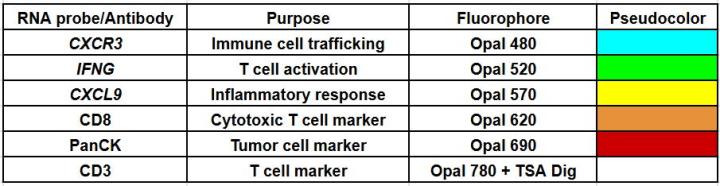
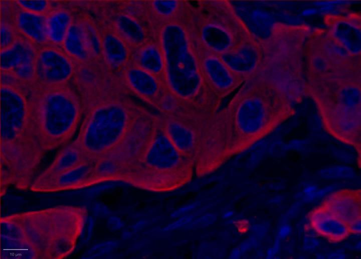
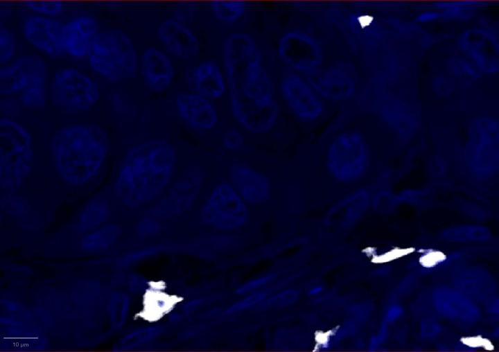
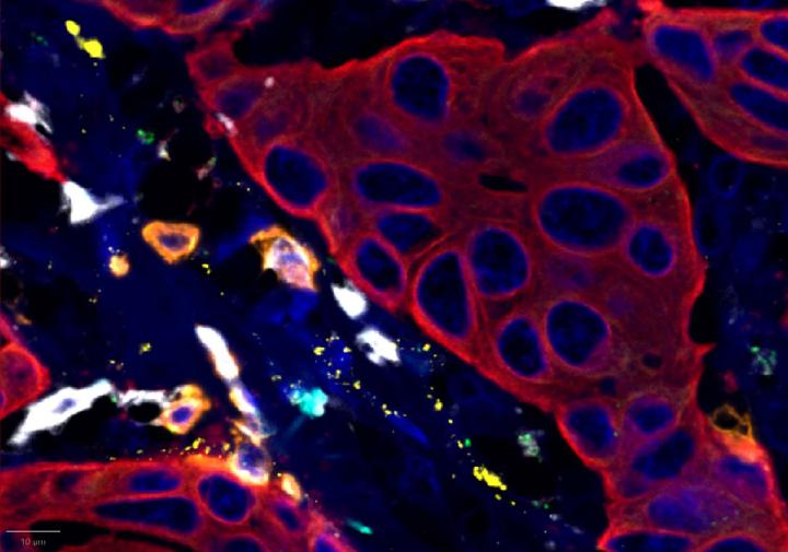
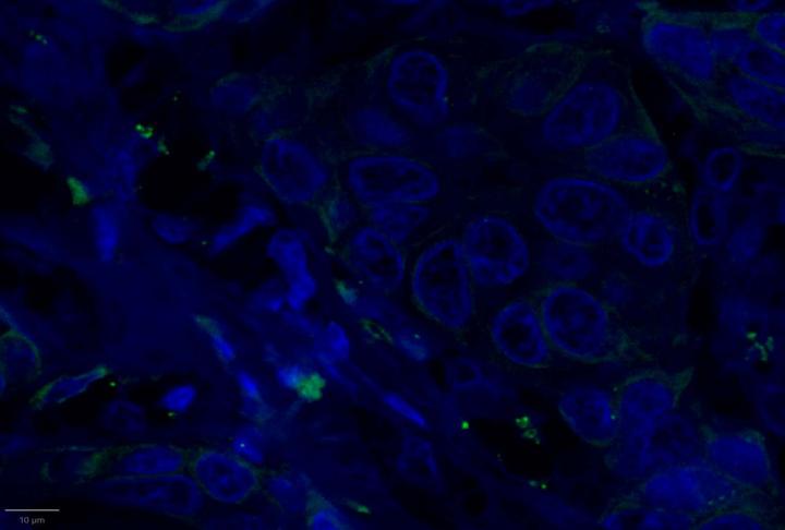
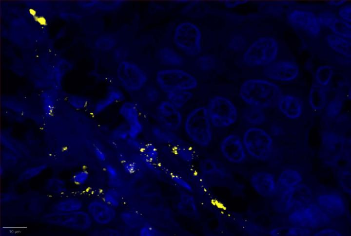
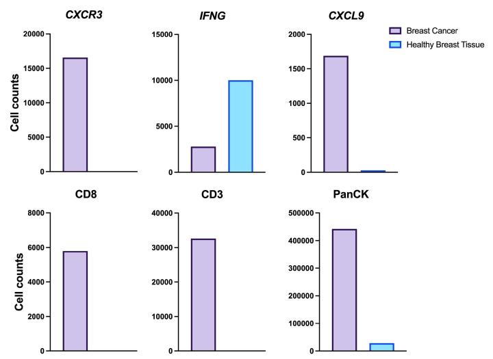
Here, we utilized RNAscope™ Multiomic LS technology to stain RNA and protein markers, CXCR3, IFNG, CXCL9, CD3, PanCK, and CD8. CXCR3 is indicative of immune cell trafficking, IFNG marks T cell activation, and CXCL9 is involved in inflammatory responses relevant to anti-tumor immunity 2-4. In conjunction, CD3 and CD8 are protein markers for T cells, and PanCK delineates tumor positive areas 5-6. Together, we have utilized a novel protease-free workflow to simultaneously detect RNA and protein markers in breast cancer tissues to further understand the tumor microenvironment.
Materials and Methods
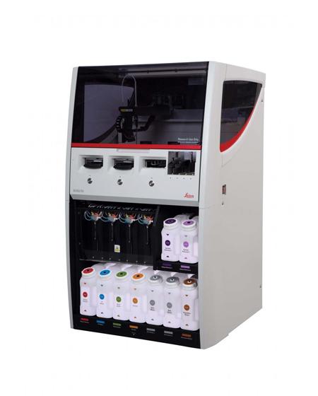
- All stains were performed using the BOND RX fully automated research stainer
- Formalin-fixed, paraffin-embedded human breast cancer and healthy breast tissue sections were used for multiomic staining.
For Research Use Only. Not for use in diagnostic procedures.
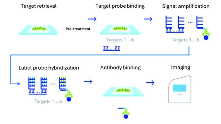
RNAscope™ Multiomic LS Assay Schematic
- Samples were imaged at 40X magnification, followed by image analysis.
- The new Multiomic LS assay supports detection of 6-plex RNA+protein targets with TSA-based detection. The workflow, run on the BOND RX, involves protease-free target retrieval followed by probe binding, signal amplification, and detection. This was followed by antibody binding for protein detection.
RNA Target, Antibody, and Fluorophore Pairings
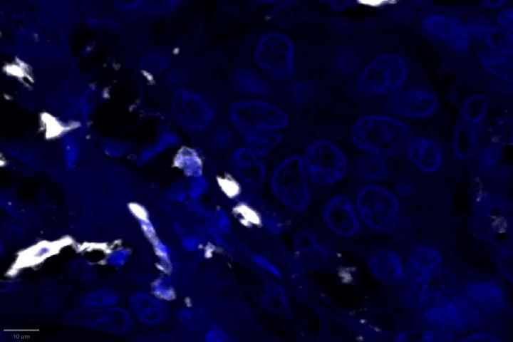
Staining Results
Positive Control Multiomic Staining in Breast Cancer
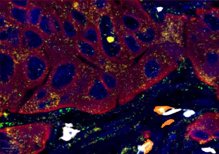
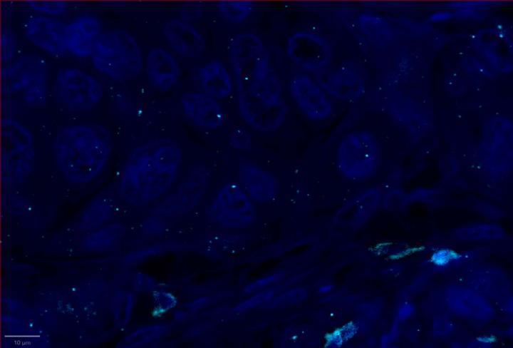
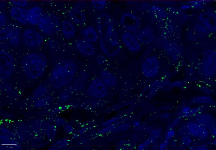
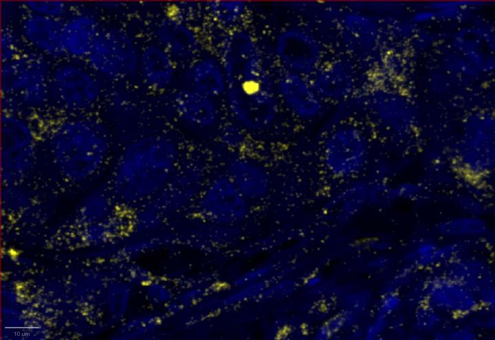
Experimental Multiomic Staining in Breast Cancer
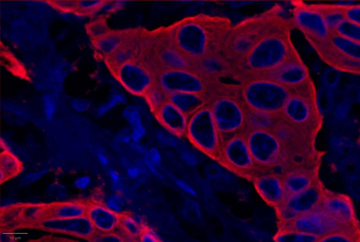
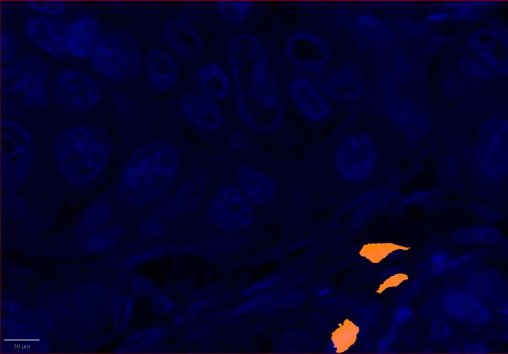
AI-Driven Image Analysis
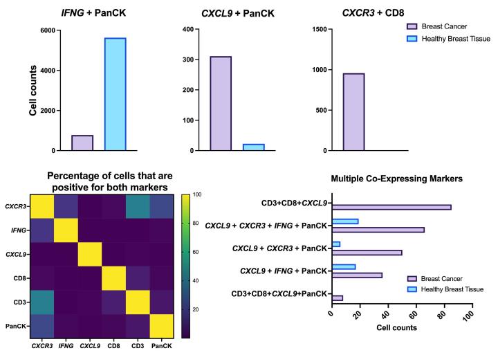
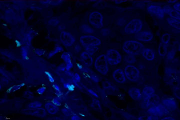
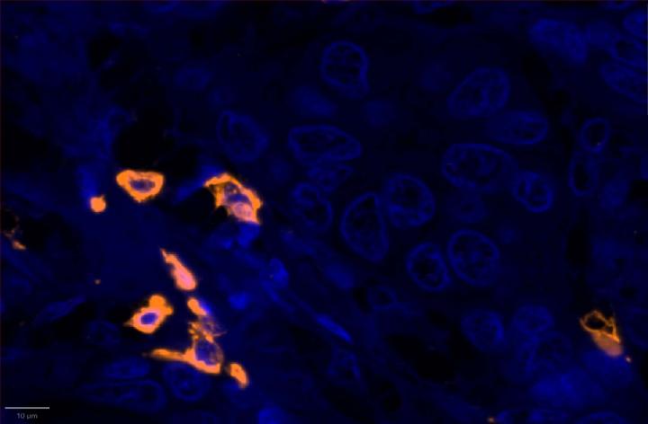
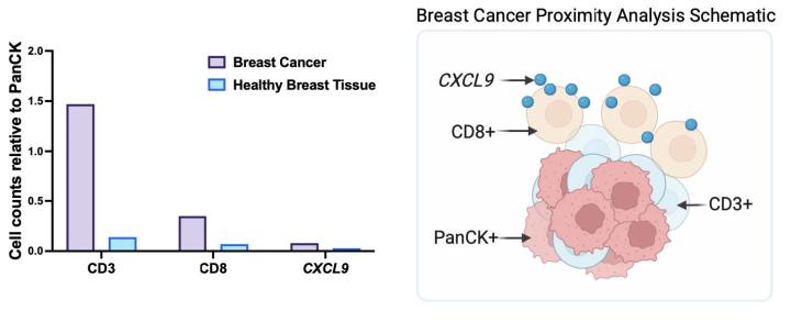
Proximity Analysis Results
Conclusions
- Compared to healthy breast tissue, the expression of CXCR3, CXCL9, CD3, CD8, and PanCK was elevated in breast cancer. This suggests strong immune cell infiltration within the tumor microenvironment. Elevated CXCR3 expression is associated with enhanced immune surveillance, while increased CXCL9 may reflect active anti-tumor immunity2,4.
- Multiomic analysis revealed co-expression of CXCR3 with CD8, indicative of T-cell recruitment7. IFNG was co-expressed with PanCK, but was down-regulated in breast cancer relative to healthy tissue, potentially indicating immune suppression and poor prognosis8.
- Proximity analysis revealed that in breast cancer, CD3, CD8, and CXCL9 were spatially adjacent to PanCK+ tumor cells, suggesting a localized pro-inflammatory response driven by cytotoxic T-cell activity9.
- The RNAscope™ Multiomic LS technology provides a powerful tool to deepen our understanding of breast cancer biology and the immune landscape of the tumor microenvironment.
Want to learn how the BOND RX can support cancer research in your lab?

참조 문헌
- Giaquinto, A. N., Sung, H., Newman, L. A., Freedman, R. A., Smith, R. A., Star, J., Jemal, A., & Siegel, R. L. (2024). Breast cancer statistics, 2024. CA: A Cancer Journal for Clinicians, 74(6), 477–495. https://doi.org/10.3322/caac.21863
- Groom, J. R., & Luster, A. D. (2011). CXCR3 in T cell function. Experimental cell research, 317(5), 620–631. https://doi.org/10.1016/j.yexcr.2010.12.017
- Jorgovanovic, D., Song, M., Wang, L., Zhang, Y. Roles of IFN-γ in tumor progression and regression: a review. Biomark Res 8, 49 (2020). https://doi.org/10.1186/s40364-020-00228-x
- Tokunaga, R., Zhang, W., Naseem, M., Puccini, A., Berger, M. D., Soni, S., McSkane, M., Baba, H., & Lenz, H. J. (2018). CXCL9, CXCL10, CXCL11/CXCR3 axis for immune activation - A target for novel cancer therapy. Cancer treatment reviews, 63, 40–47. https://doi.org/10.1016/j.ctrv.2017.11.007
- Sun X, Zhai J, Sun B, Parra ER, Jiang M, Ma W, Wang J, Kang AM, Kannan K, Pandurengan R, Zhang S, Solis LM, Haymaker CL, Raso MG, Mendoza Perez J, Sahin AA, Wistuba II, Yam C, Litton JK, Yang F. Effector memory cytotoxic CD3+/CD8+/CD45RO+ T cells are predictive of good survival and a lower risk of recurrence in triple-negative breast cancer. Mod Pathol. 2022 May;35(5):601-608. doi: 10.1038/s41379-021-00973-w. Epub 2021 Nov 27. PMID: 34839351.
- Ji H, Yuan L, Jiang Y, Ye M, Liu Z, Xia X, Qin C, Jiang D, Gai Y, Lan X. Visualizing Cytokeratin-14 Levels in Basal-Like Breast Cancer via ImmunoSPECT Imaging. Mol Pharm. 2022 Oct 3;19(10):3542-3550. doi: 10.1021/acs.molpharmaceut.2c00004. Epub 2022 Mar 14. PMID: 35285645.
- Kurachi, M., Kurachi, J., Suenaga, F., Tsukui, T., Abe, J., Ueha, S., Tomura, M., Sugihara, K., Takamura, S., Kakimi, K., & Matsushima, K. (2011). Chemokine receptor CXCR3 facilitates CD8(+) T cell differentiation into short-lived effector cells leading to memory degeneration. The Journal of experimental medicine, 208(8), 1605–1620. https://doi.org/10.1084/jem.20102101
- Lamsal, A., Andersen, S. B., Johansson, I., Vietri, M., Bokil, A. A., Kurganovs, N. J., Rylander, F., Bjørkøy, G., Pettersen, K., & Giambelluca, M. S. (2023). Opposite and dynamic regulation of the interferon response in metastatic and non-metastatic breast cancer. Cell communication and signaling : CCS, 21(1), 50. https://doi.org/10.1186/s12964-023-01062-y
- Liang, H., Huang, J., Li, H., He, W., Ao, X., Xie, Z., Chen, Y., Lv, Z., Zhang, L., Zhong, Y., Tan, X., Han, G., Zhou, J., Qiu, N., Jiang, M., Xia, H., Zhan, Y., Jiao, L., Ma, J., Radisky, D., … Zhang, X. (2025). Spatial proximity of CD8+ T cells to tumor cells predicts neoadjuvant therapy efficacy in breast cancer. NPJ breast cancer, 11(1), 13. https://doi.org/10.1038/s41523-025-00728-9
Related Content


Leica Biosystems 콘텐츠는 Leica Biosystems 웹사이트 이용 약관의 적용을 받으며, 이용 약관은 다음에서 확인할 수 있습니다. 법적고지. 라이카 바이오시스템즈 웨비나, 교육 프레젠테이션 및 관련 자료는 특별 주제 관련 일반 정보를 제공하지만 의료, 규정 또는 법률 상담으로 제공되지 않으며 해석되어서는 안 됩니다. 관점과 의견은 발표자/저자의 개인 관점과 의견이며 라이카 바이오시스템즈, 그 직원 또는 대행사의 관점이나 의견을 나타내거나 반영하지 않습니다. 제3자 자원 또는 콘텐츠에 대한 액세스를 제공하는 콘텐츠에 포함된 모든 링크는 오직 편의를 위해 제공됩니다.
모든 제품 사용에 다양한 제품 및 장치의 제품 정보 가이드, 부속 문서 및 작동 설명서를 참조해야 합니다.
Copyright © 2026 Leica Biosystems division of Leica Microsystems, Inc. and its Leica Biosystems affiliates. All rights reserved. LEICA and the Leica Logo are registered trademarks of Leica Microsystems IR GmbH.