
Digital Pathology in Toxicological Pathology Studies

Drug development is a timely and costly process, 1 with the average cost of bringing a single new drug to market costing $1.3 billion, 2 taking anywhere between 10-15 years to finalize complete all clinical trials. 3 Preclinical studies are the vital first steps in research and development and is estimated to cost $7M over 73 months. 4 Up to 35% of drugs are eliminated at phase I and II clinical trials due to toxicity. 5
Toxicological pathologists are vital to the drug discovery process to identify efficacious and toxic effects of new drugs by histopathological analysis of animal model tissues that determines the no adverse effect level that will be used for phase 1 clinical trials. 6
Peer review is commonplace for toxicology pathology to solidify and enhance scientific accuracy and quality of the findings. 7 These findings could determine whether the compound should be pursued or terminated based on safety and efficacy. 6 It is important to make such decisions early in the drug discovery process to improve the return on investment for pharmaceutical companies. 6
However, many toxicological pathologists are faced with an inefficient workflow that prolongs the time for peer review due to slide packing, delivery, annotation etc., as well as geography of collaborators. The diagram below depicts the conventional peer review process.

Digital pathology can help to eliminate this workflow challenge when implemented into the toxicology pathology workstream. Digital pathology is the process of scanning whole slides to produce a digital image of the tissue that can be sorted, annotated, and shared virtually on a computer or mobile device. Digital pathology produces images at the same quality and resolution as the conventional microscope. Below is how the manual workflow can be changed with digital pathology:

In toxicological studies, additional advantages of digital pathology are becoming more widely recognized. This includes, but is not limited to:
- Reduce duplication of annotations and reports. 1
- Compare multiple slides side by side for analysis. 1
- Use metadata or organize, select, and sort slides for review within designated software. 1,8
- Reduce cost of pathologist’s travel and slide shipping, as well as reducing risk of damage to fragile slides. 8
- Standardize pathology and training for consistency of interpretation. 1
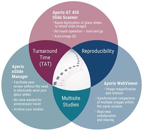
Leica Biosystems offers Aperio Digital Pathology solutions for whole slide scanning, web viewing and data management. This is highlighted in the diagram below:
발표자 소개
Rhian is a Scientist from Swansea University in Medical and Healthcare Studies and was featured in several collaborative publications. Rhian’s research-based background focused on tissue-based pathology in Multiple Sclerosis, primarily using immunohistochemical analysis and in vitro molecular techniques. She spent a short period conducting routine PCR testing for COVID-19 at the end of 2020.
참조 문헌
- Potts SJ. Digital pathology in drug discovery and development: multisite integration. Drug discovery today. 2009;14(19/20): 935-941.
- Wouters OJ, McKee M, Luyten J. Estimated Research and Development Investment Needed to Bring a New Medicine to Market, 2009-2018. JAMA. 2020;323(9):844–853. doi:10.1001/jama.2020.1166
- Cancer Research UK. How long a new drug takes to go through clinical trials. Cancer Research UK website. February 22, 2019. Accessed May 6, 2021. www.cancerresearchuk.org/find-a-clinical-trial/how-clinical-trials-are-planned-and-organised/how-long-it-takes-for-a-new-drug-to-go-through-clinical-trials
- Vieira M. Research synthesis: Costs of Pharmaceutical R&D. The Graduate Institute of Geneva website. January, 2020. Accessed May 6, 2021. da7af2c8-d9b0-47a3-a3f6-89c3c3bfa02c.filesusr.com/ugd/356854_e9d75e29c0264bf9b38118fc5f0aeab6.pdf DocuSign Envelope ID: FCD13E60-AFFE-41BF-856D-D9A6D04254EB
- Donowitz M, Turner JR, Verkman AS, Zacho NC. Current and potential future applications of human stem cell models in drug development. J Clin Invest. 2020;130(7):3342-3344.
- van Tongeren S, Fagerland JA, Conner MW, et al. The Role of the Toxicologic Pathologist in the Biopharmaceutical Industry. International Journal of Toxicology. 2011;30(5):568-582. doi:10.1177/1091581811413304
- Morton D, Sellers RS, Barale-Thomas E, Bolon B, George C, Hardisty JF, Irizarry A, McKay JS, Odin M, Teranishi M. Recommendations for pathology peer review. Toxicol Pathol. 2010 Dec;38(7):1118-27. doi: 10.1177/0192623310383991. Epub 2010 Oct 5. PMID: 20924082.
- Hamilton PW, Bankhead P, Wang Y, et al. Digital pathology and image analysis in tissue biomarker research. Methods. 2014;70(1):59-73. doi.org/10.1016/j.ymeth.2014.06.015
Related Content


Leica Biosystems 콘텐츠는 Leica Biosystems 웹사이트 이용 약관의 적용을 받으며, 이용 약관은 다음에서 확인할 수 있습니다. 법적고지. 라이카 바이오시스템즈 웨비나, 교육 프레젠테이션 및 관련 자료는 특별 주제 관련 일반 정보를 제공하지만 의료, 규정 또는 법률 상담으로 제공되지 않으며 해석되어서는 안 됩니다. 관점과 의견은 발표자/저자의 개인 관점과 의견이며 라이카 바이오시스템즈, 그 직원 또는 대행사의 관점이나 의견을 나타내거나 반영하지 않습니다. 제3자 자원 또는 콘텐츠에 대한 액세스를 제공하는 콘텐츠에 포함된 모든 링크는 오직 편의를 위해 제공됩니다.
모든 제품 사용에 다양한 제품 및 장치의 제품 정보 가이드, 부속 문서 및 작동 설명서를 참조해야 합니다.
Copyright © 2026 Leica Biosystems division of Leica Microsystems, Inc. and its Leica Biosystems affiliates. All rights reserved. LEICA and the Leica Logo are registered trademarks of Leica Microsystems IR GmbH.