Breast Ki67 AI
Developed by Indica Labs, the Breast Ki67 AI solution leverages deep learning that standardizes scoring of Ki67 in resections, excisions, or biopsies from primary invasive breast cancer samples. The algorithm reports quantitative results and markup images, including tumor detection and percent positive Ki67 cells.
Breast Ki67 AI is For Research Use Only and not intended for clinical diagnostic use. Breast Ki67 AI accessed via the Aperio HALO AP image management system.

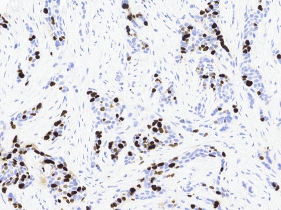
Step 1: Breast Ki67 IHC Image
Breast Ki67 AI standardizes quantification of Ki67 IHC-stained breast cancer resections, excisions and biopsy samples.
The algorithm can be launched by a user directly in the viewer or configured to automatically run for every breast Ki67 image in Aperio HALO AP.
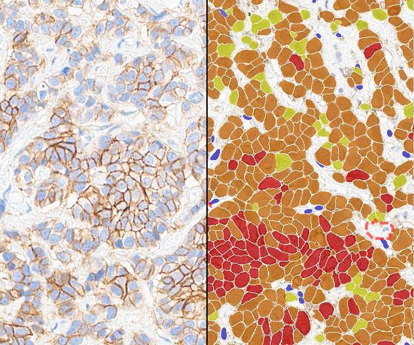
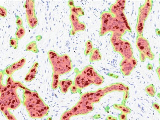
Step 2: Breast Ki67 IHC Tumor Detection
An image analysis heatmap mask is generated, denoting regions within the sample containing invasive breast cancer.
Prior to this step, image analysis pre-processing excludes artifact and benign epithelial tissue.
The following metrics are calculated as outputs for each slide:
- Tumor Area
- Other Area
- % Tumor Area
- Classified Area
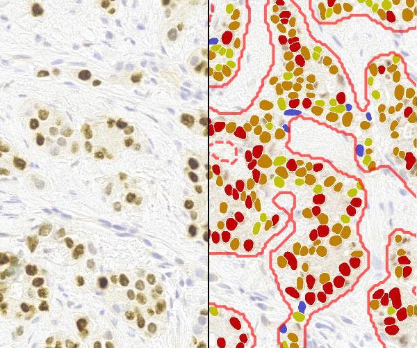
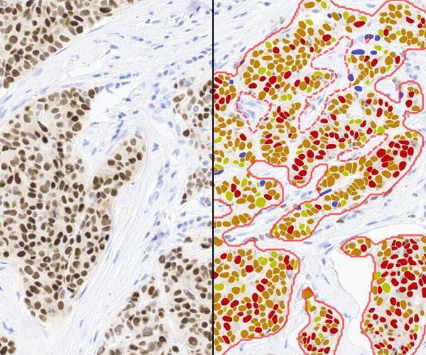
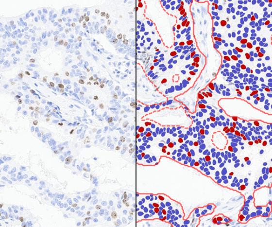
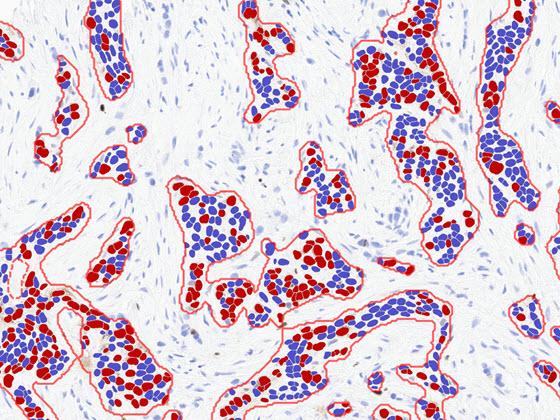
Step 3: Ki67 Cell Classification
Breast Ki67 AI generates an image mask segmenting cells within the tumor region, denoting the following cell classes:
- Negative Ki67 Cell: Blue
- Positive Ki67 Cell: Red
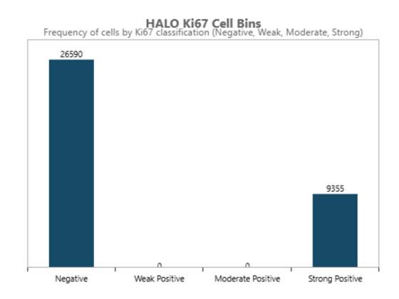
Results: Histogram
Breast Ki67 AI generates a histogram displaying the total number of cancer cells classified within the following categories:
- Ki67 Negative
- Ki67 Positive (Strong)
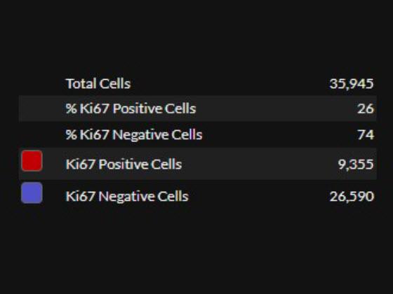
Results: Quantitative
Breast Ki67 AI generates the following metrics as output for the whole slide image:
- Total Cells
- % Ki67 Positive Cells
- % Ki67 Negative Cells
- Number of Ki67 Positive Cells
- Number of Ki67 Negative Cells
Breast Ki67 AI in Aperio HALO AP Demonstration
Explore the future of pathology with the Breast Ki67 AI app from Indica Labs. Breast Ki67 AI seamlessly integrates within the Aperio HALO AP enterprise digital pathology platform to identify Ki67 tumor cells in primary invasive breast cancer samples.
After selecting an image, view the IHC slide in the viewer and the Breast Ki67 AI results. Results include overlays and quantitative results. The first set of overlays prepare the tissue for analysis by identifying areas of analyzable tissue. The next overlay identifies areas of tumor in the tissue. The final overlay identifies and classifies cells for Ki67 positivity within the tumor area. Finally, quantitative results at a per slide level are displayed in the assay panel to the right.
Documents
Related Product Applications