
The Future is Now: Digital Pathology is Pathology
In April 2021, the Digital Pathology Association (DPA), European Society of Digital and Integrative Pathology (ESDIP) and Japanese Society of Digital Pathology (JSDP) established a global task force with the intent of promoting worldwide adoption of digital pathology. This alliance aims to boost the exchange of knowledge across borders, unify concepts and standardization, and help break down political, social, and economic barriers, including things as basic as mistrust in digital pathology.1
Simply stated, digital pathology is the present. It's not the future anymore. As we and the other global task force members have noted2, the time to strike is now when the current technological and regulatory climate is favorable to support the digital transformation of pathology. Mature digital imaging technology exists to support a digital transformation in pathology, but transformation remains hindered by skepticism about the viability of adopting a digital workflow in routine clinical practice.1 Worldwide collaboration is needed to address the skepticism, to recognize we have commonalities – such as a shortage of pathologists and economic pressures – and to acknowledge important regional differences such as local regulatory requirements. We have a winning situation for everybody when we operate together to understand the difficulties in different countries, in different continents and to try to accelerate and remove some myths around digital pathology.
Recently, we participated in a webinar regarding digital transformation moderated by Colin White of Leica Biosystems and hosted by The Pathologist. We are encouraged by the strong response to the session, which attracted more than a thousand registrants, and the thoughtful questions from participants regarding how to start a journey to digital. The following principles emerged as foundational to digital transformation:
We must change the way pathologists think about the pathology lab and methodologies to address current and future demands, versus romanticizing how pathologists used to process samples. Education will be vital in this endeavor. In Japan, for example, Dr. Fukuoka’s fellows don't start with a microscope, so they don't need any special education in digital pathology. Experienced pathologists should be made aware of the tremendous progress that has been made to address historic challenges to digital pathology adoption. Many of the historic technology, cost and regulatory barriers have been overcome. We have cloud computing. We have infrastructure. Furthermore, we are seeing a reduction in the cost of digitizing per slide. But even with all these advancements, pathologists are still hesitant to adopt digital pathology.
To overcome pathologists’ hesitance, it is helpful to start the journey to digital sooner rather than waiting, and to start small. Our collective experience is to focus at the outset on building something good and demonstrating that it's going to solve a gap in current practice. People will use it and you can grow from there. At the start of implementation expect a few users, whose experience will resonate among and attract other pathologists to see digital pathology in action. From there, we’ve found adoption will exponentially grow.
Once a pathologist is presented with the perfect whole slide image, there is no way he/she will want to go back to the microscope. You’ll achieve a double goal: make pathologists happy and prepare yourself for AI. We’ve found that the usage of the whole slide images invites exploration of AI and predicts parallel growth. The potential of AI is exciting, and ushers in the possibility of working synergy with AI tools and having reports which have more quantified data. Ten years ago, we used to talk about the promise of AI. AI is not a promise anymore. It's a reality. The first AI app has FDA clearance in the United States. There are many more which are on the verge of being released. There is commercially available software that pathologists can use and there is also open-source software available. We’re shifting to an era where there will be no digital pathology or AI, it's all pathology.
We welcome pathologists and the broader pathology community worldwide to listen to the replay of the webinar and join future Global Task Force events.
About the presenters
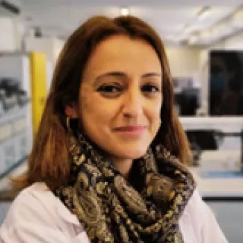
Dr. Catarina Eloy is the Head of the Pathology Laboratory of IPATIMUP/ Researcher at i3S/ Affiliated Professor of Medical Faculty of The University of Porto. She is also the former President of the European Society for Digital and Integrative Pathology and Vice-president of the Portuguese Society of Anatomic Pathology.
Dr. Eloy received her MD in 2003 from the Medical Faculty of The University of Porto with a specialization in Pathology
Dr. Eloy has been a researcher since 2006 at the cancer signaling & metabolism of i3S/Ipatimup and received a Ph.D. in Thyroid cancer in 2012 by the Medical Faculty of The University of Porto.
Dr. Eloy has 70 publications and three academic and scientific awards/distinctions.
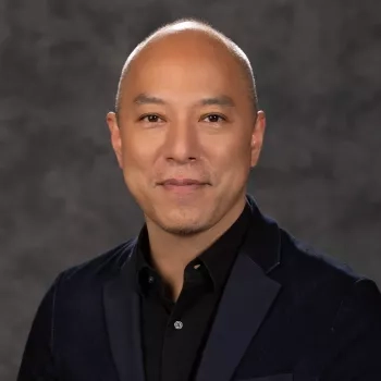
Prof. Jun Fukuoka is a Japanese pathologist whose expertise lies in the field of pulmonary disease and digital pathology. He has graduated from Shiga University of Medical Science in 1995 and studied lung pathology and molecular research at Mayo Clinic, AFIP, and NIH.
He was promoted to full professor in 2009 in Toyama University and relocated as a department chair and professor at Nagasaki University in 2012. From 2017, he started cross-appointment with one of the largest hospitals, Kameda Medical Center, Department of Pathology, in Japan. He allocates 60% of his time at Nagasaki University and 40% at Kameda Medical Center.
He has launched two start-up companies during his career in the field of pathology. He is also a good teacher and has developed more than 30 pathologists worldwide and has mentored more than 100 undergraduate students/residents on either presenting research works in international meetings or publishing in scientific journals.
He has started research in digital pathology since 2010 and has developed a fully digitized network to share diagnostic process and education among several large institutes. Through the network, virtual large laboratory among several institutes has been created. His young team is enthusiastic to develop the workflow of human-AI collaboration.
He currently serves as Vice-President at Japanese Society of Digital Pathology (JSDP) and Treasurer of Digital Pathology Association (DPA).
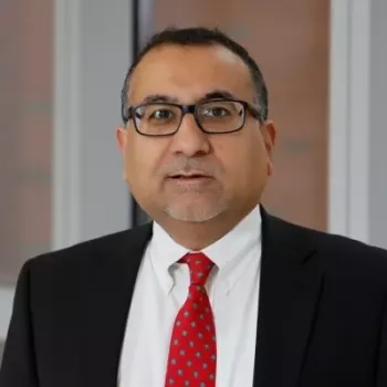
Dr. Anil Parwani is a Professor of Pathology and Biomedical Informatics at The Ohio State University. He serves as the Donald A. Senhauser Chair of the Department of Pathology and the Chief of Pathology Services for the Health System.
Dr. Parwani has expertise in surgical pathology, pathology informatics, whole slide imaging, telepathology, image analysis, artificial intelligence, and lab automation. Dr Parwani has authored over four hundred peer-reviewed articles in major scientific journals and several books and book chapters.
Dr. Parwani has served on the board of directors, education committee and abstract committee for USCAP and was the president of digital pathology association. Dr. Parwani is the Editor-in-chief of Diagnostic Pathology and Co-editor of the Journal of Pathology Informatics.
References
- Eloy C, Bychkov A, Pantanowitz L, Fraggetta F, Bui MM, Fukuoka J, Zerbe N, Hassell L, Parwani A. DPA–ESDIP–JSDP task force for worldwide adoption of digital pathology. J Pathol Inform 2021;12:51.
- Griffin J, Treanor D. Digital pathology in clinical use: Where are we now and what is holding us back? Histopathology 2017;70:134-45.
Related Content


Leica Biosystems Knowledge Pathway content is subject to the Leica Biosystems website terms of use, available at: Legal Notice. The content, including webinars, training presentations and related materials is intended to provide general information regarding particular subjects of interest to health care professionals and is not intended to be, and should not be construed as, medical, regulatory or legal advice. The views and opinions expressed in any third-party content reflect the personal views and opinions of the speaker(s)/author(s) and do not necessarily represent or reflect the views or opinions of Leica Biosystems, its employees or agents. Any links contained in the content which provides access to third party resources or content is provided for convenience only.
For the use of any product, the applicable product documentation, including information guides, inserts and operation manuals should be consulted.
Copyright © 2026 Leica Biosystems division of Leica Microsystems, Inc. and its Leica Biosystems affiliates. All rights reserved. LEICA and the Leica Logo are registered trademarks of Leica Microsystems IR GmbH.