52 result(s) for 'Tissue Preparation'
31 - 40 of 52 results for 'Tissue Preparation'
Sort by
Show
Skin specimens received in the histology laboratory for dermatopathology are among the most difficult to handle successfully. The pathologist must be able to see the dermal-epidermal junction in each tissue section in order to make a diagnosis, thus every skin...

Different types of cancers frequently metastase to bone tissue. Treatment planning decisions are often based upon histology and special staining of these distant sites of disease. These decisions may rely on the outcome of immunohistochemistry, in situ...

This presentation will review the challenges of creating a system to assess the quality and consistency of diagnoses produced by individual pathologists and discuss setting goals to improve the processes in anatomic pathology for enhanced patient safety. All...

When was the last time that your pathologist brought you a slide of decalcified bone, and said it was the best she ever saw? Ever wonder why your PAS stain is not staining the basement membrane the way it should? These questions and 18 others will be discussed...

Producing an H&E stained slide is a process. It starts way before the slide is loaded on a stainer or moved down through containers by hand. Producing quality, consistent and reproducible H&E stained slides is a process as well as a lesson in...

In recent years, there has been an increase of pathogens findings in tissues. This presentation will discuss some of the reasons for the increase of these pathogens in the past few years. The presentation will also will give an overview of why some of these...
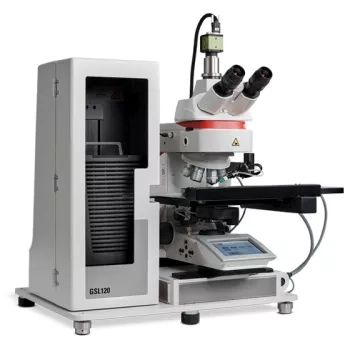
Flexible Image Analysis Platform for Every Cytogenomics Laboratory
The CytoInsight GSL automated image analysis and case management system is a comprehensive Cytogenomics solution that improves quality and turnaround time from acquisition to analysis while reducing hands-on time at every step in your process.
The CytoInsight GSL system improves image processing, provides system flexibility, and offers increased cybersecurity to deliver the tools to build your system to fit your Cytogenomics lab workflow.
From prepared slide to report and back, the CytoInsight GSL flexible platform for brightfield and fluorescence image analysis provides unlimited scanning for a walk-away solution to meet the evolving needs of your Cytogenomics laboratory.

Slide 1
Hello, hello; thank you so much for that introduction. Let's get started. Oh, I'm sorry, I just need to hit this next button here. So my name is Mark Lawson.
Thank you for joining us in this webinar. I'm here to present BOND RX tips, tricks, and optimization. It's going to be a user guide from the BOND RX and chromogenic multiplexing in the research application.
Slide 2
So, my name is Mark Lawson. I'm an application specialist on the Life Sciences team at Leica Biosystems I provide technical support for the Life Sciences portfolio, including but not limited to the BOND RX, the BOND RXm, and a wide array of reagents. So I've worked in the histology field for about 15 years and in both clinical and research spaces. I started off as a histotechnologist and worked my way up...

One of the most critical steps in histology is fixation, especially when it comes to fatty tissue.

In recent years, digital pathology has gone from an intriguing idea to an integral part of how academic and commercial labs operate. Join Leica Biosystems and Procia Digital Pathology as they discuss why institutions are going digital today and how they...