52 result(s) for 'Tissue Preparation'
21 - 30 of 52 results for 'Tissue Preparation'
Sort by
Show
BOND ancillaries have been designed for Leica Biosystems fully-automated BOND systems: BOND-MAX and BOND-III systems. Used in the development and validation of BOND ready-to-use primary
antibodies and ISH probes - choose Leica Biosystems reagents for the assurance of a quality result in your laboratory. BOND Wash Solution 10X Concentrate is a concentrated buffer solution, requiring initial
dilution. The diluted solution is for washing sections of...
Antigen Background
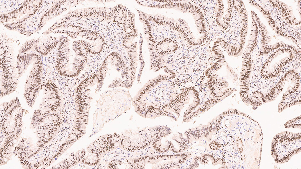
Postmeiotic segregation increased 2 (PMS2), also known as PMS1 protein homologue 2, is a DNA mismatch repair (MMR) protein. The PMS2 gene family members are found in clusters on chromosome 7. PMS2 is a 96 kDa mismatch repair protein closely related to MLH1, MLH3 and PMS1, which are homologs of the bacterial mutL gene. The PMS2 protein forms a heterodimer with the MLH1 protein which is then activated in the presence of ATP; this...
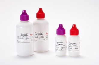
“Novolink Polymer Detection Systems are used for the visualization of mouse IgG, mouse IgM and rabbit IgG primary antibodies. Novolink Polymer contains component reagents of these systems. The
Novolink Polymer Detection Systems utilize a novel controlled polymerization technology to prepare polymeric HRP-linker antibody conjugates. Therefore, the problem of non-specific staining that can occur with Streptavidin/Biotin detection systems due
to end...


In surgical pathology more than ever, the laboratories are expected to do more with less. Taking on more specimens with less technical staff, or getting more stains out of tiny biopsies or fine needle aspirations have become the norm. The College of American...
Integrating Innovation with Automation for the Research Laboratory
At Leica Biosystems, we aim to enable researchers to accelerate their journey, transforming scientific exploration into translational outcomes. Our goal is to shape the future with novel technologies that inspire every researcher’s exploration of biology.
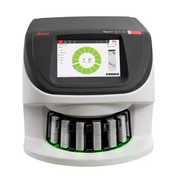
The Aperio GT 450 v 1.3 is proven scanning technology that consists of three upgrades that improve image quality and flexible scanning solutions in research settings: Z-Stack Scanning, Automatic Narrow Stripe, and 20x Magnification.
Z-Stack Scanning
Multiplane scanning that produces a composite, 3D image that enables research pathologists to review slide samples at varied levels of thickness, similar to a traditional microscope.
Automatic Narrow Stripe Scanning...

One of the most fundamentally critical elements of diagnostic histopathology is first the ability to suspend all cellular activity in tissue and prevent degradation, and secondly to process that specimen in a manner that facilitates subsequent steps such as...

A review of fluorochromes and the specialized microscope used in immunofluorescence techniques will begin this presentation. A typical (human skin and kidney) clinical specimen will be followed from receipt in the laboratory, through freezing, cryomicrotomy...

The potential for non-invasive tests that provide equivalent research and diagnostic value as can be obtained from tissue biopsies is real, but not yet realized. Tissue biopsies allow for identification, phenotyping and molecular analysis of cancer and...

In this session, we will briefly review the basics of molecular biology, examine critical factors which affect the quality of nucleic acids in the tissues and cells which are submitted for downstream molecular diagnostics, and briefly introduce some...