57 result(s) for 'Tissue Preparation'
1 - 10 of 57 results for 'Tissue Preparation'
Sort by
Show
*/
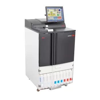
This Item has been discontinued
Replaced by HistoCore PEGASUS
Total Confidence in Every Tissue
Boost Your Confidence in Quality and Reliable Tissue Processing
Pathology laboratories are a busy place and there are many challenges to think about. To give you peace of mind, make sure to choose a tissue processor that lets you focus on the important things. The ASP6025 S tissue processor supports your laboratory with the following features:
An advanced magnetic stirrer technology optimizes paraffin infiltration and enhances reagent exchange within the tissue cells.
Density Meter tracks reagent concentration for improved processing quality and reduced user errors.
Pre-installed validated protocols give you the flexibility to...
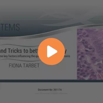
In this webinar, Fiona Tarbet will investigate some of the effects of poor technique on section and stain quality and identify ways of producing better results.
Slide 1
Welcome
Slide 2
Slide 3
Discuss Pre-Analytics and the impact of improper fixation and artifacts on tissue processing
Discuss Fixation and the impact of incomplete fixation on tissue processing
Describe the impact of improper Prosection
Determine satisfactory Processing of samples
Explain why routine Maintenance is a critical success factor to proper tissue processing
Slide 4
This slide has two examples of what the ideal sections in a perfect world look like under the scope. The skin section on the left is clean looking with the Eosin staining the components of the dermis with different shades. The colon section on the right is crisp with well-defined nuclei and cilia. There is no background staining or muddiness to the stain.
Slide 5
One artifact seen the lab that is n...

Learn more about tissue processing artifacts, what is "optimal" and what is "not," and how to optimize tissue processing techniques with a downloadable checklist for success.
Following our two-part webinar series of Tips & Tricks to Better Histology, further questions were raised by customers referring to specific issues encountered during histology/staining practice. Some of these questions are answered here to help combat issues experienced in the laboratory.
Following our 2-part webinar series of Tips & Tricks to Better Histology, questions about histology-related issues were received from customers and answered by Leica Biosystems. Here, in Histology Tips & Tricks: Questions and Answers, Part 2, more questions are answered, specifically regarding decalcifying agents, and tissue section bubbling/cracking.
Slide 1
Welcome!
Slide 2
Intro slide
Slide 3
It's nice to meet you!
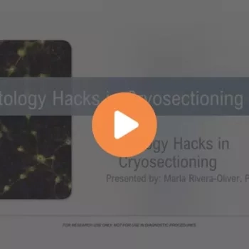
Welcome to the Histology Hacks in Cryosectioning webinar.
I am Dr. Marla Rivera-Oliver, and I am very excited to share today some tips and tricks I've learned during my ten years of experience working on basic and translational research. I am based in the Dallas, Texas, area of the United States, and in my current role, I support customers from North Texas and Oklahoma. I completed a bachelor's in biology from the Interamerican University of Puerto Rico and a doctoral degree in Neurobiology from the University of Puerto Rico. I specialized in the spinal cord injury field and honed my skills in Cryosectioning, imaging, and in vivo electrophysiology. Then, I moved into a post-doctoral position at the Center for D...