10 result(s) for 'Histology'
1 - 10 of 10 results for 'Histology'
Sort by
Show
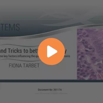
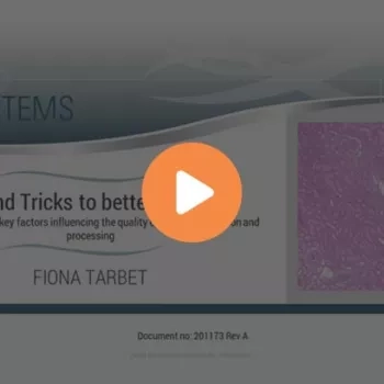
Following our two-part webinar series of Tips & Tricks to Better Histology, further questions were raised by customers referring to specific issues encountered during histology/staining practice. Some of these questions are answered here to help combat issues experienced in the laboratory.
Following our 2-part webinar series of Tips & Tricks to Better Histology, questions about histology-related issues were received from customers and answered by Leica Biosystems. Here, in Histology Tips & Tricks: Questions and Answers, Part 2, more questions are answered, specifically regarding decalcifying agents, and tissue section bubbling/cracking.
Slide 1
Welcome
Slide 2
Slide 3
Discuss Pre-Analytics and the impact of improper fixation and artifacts on tissue processing
Discuss Fixation and the impact of incomplete fixation on tissue processing
Describe the impact of improper Prosection
Determine satisfactory Processing of samples
Explain why routine Maintenance is a critical success factor to proper tissue processing
Slide 4
This slide has two examples of what the ideal sections in a perfect world look like under the scope. The skin section on the left is clean looking with the Eosin staining the components of the dermis with different shades. The colon section on the right is crisp with well-defined nuclei and cilia. There is no background staining or muddiness to the stain.
Slide 5
One artifact seen the lab that is n...


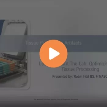
Learn more about tissue processing artifacts, what is "optimal" and what is "not," and how to optimize tissue processing techniques with a downloadable checklist for success.
Digital pathology is a growing field, with multiple vendors offering a variety of hardware and software for different applications. With many options available for digital pathology scanners, viewing software, and both consumer and medical review monitors, users need the ability to adapt to variations in image appearance.

Digital pathology is quickly growing across the globe as it offers increasing benefits to standardize and optimize the pathology lab workflow. Pathologists can now retrieve their cases in digital form and review and sign off cases using a PC and monitor. However, digital pathology companies offer many different monitors, depending on the company. Pathologists are often frustrated by the color variation of a digital image as it may not match the microscope color experience They often prefer a color that looks as close as possible to the typical experience using a microscope.