



CytoVision DX Automated Cytogenetics Imaging, Analysis, and Case Management System


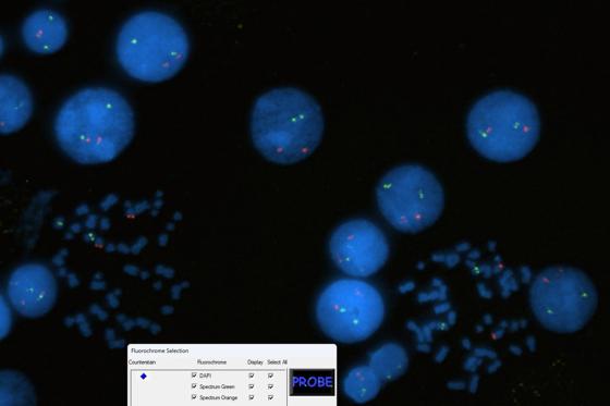
Overview
Proven Precision and Performance
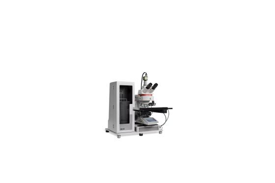
CytoVision DX is a fully automated brightfield and fluorescence system for image capture, analysis, and case management in clinical cytogenetics laboratories. It supports karyotyping, interphase FISH, tissue FISH, m-FISH, and related cytogenetic applications in a connected workflow from slide to report. Developed with leading cytogenetics laboratories and built on more than 30 years of CytoVision performance, the system combines automated sample capture, high-resolution imaging, analysis software, and case management.
For In Vitro Diagnostic Use.
Features
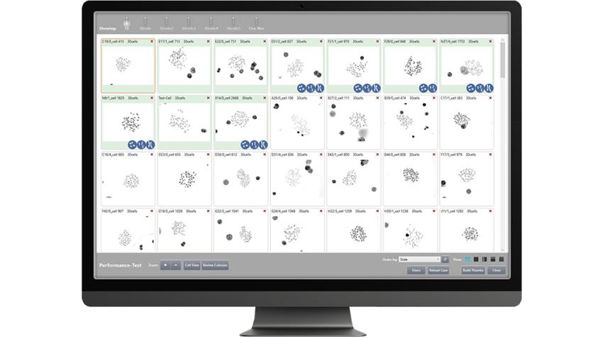
Automated Cytogenetics Imaging, Analysis, and Case Management
CytoVision DX combines image capture, analysis, and case management capabilities within a single platform designed for clinical cytogenetics laboratories. Supporting karyotyping, interphase FISH, tissue FISH, m-FISH, and spot-counting applications, the system helps streamline workflows from image acquisition through case review and reporting.
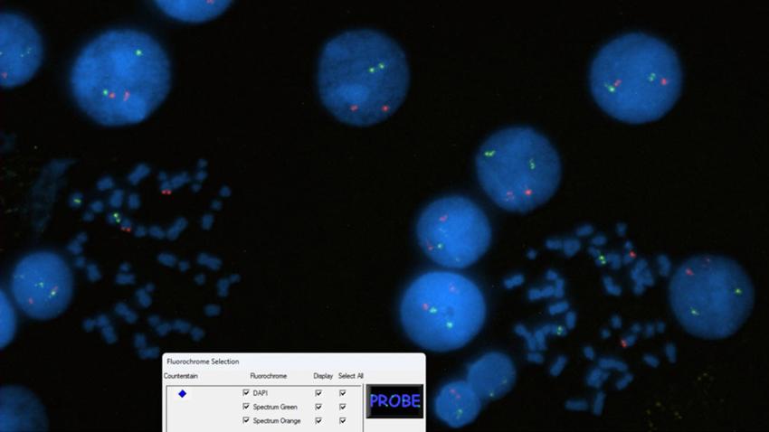
Automate Cell Identification, Capture, and Analysis Workflows
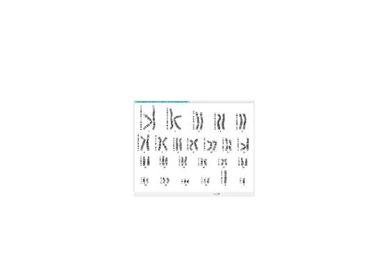
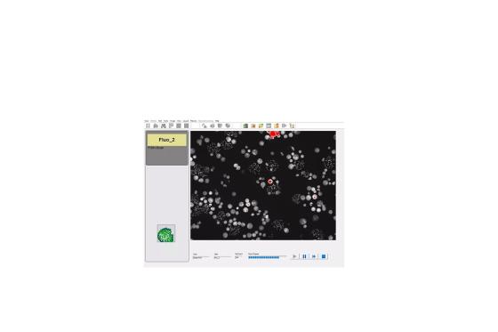
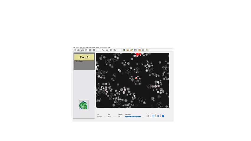
CytoVision DX automatically identifies, locates, and captures metaphase and interphase cells to reduce manual image acquisition steps. The platform also supports karyogram creation and automates many routine imaging tasks, helping laboratory personnel focus more time on analysis and interpretation. Mixed-batch scanning allows karyotyping, interphase FISH, and tissue FISH specimens to be processed within a single workflow, improving flexibility and operational efficiency.
Support Karyotyping, FISH, and Advanced Cytogenetics Applications
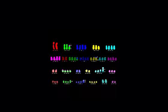
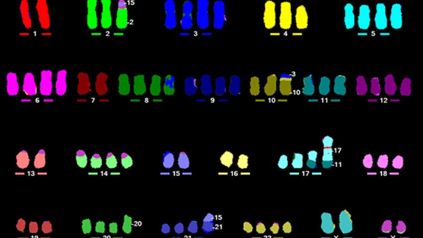
A single platform supports multiple clinical cytogenetics workflows, including karyotyping, interphase FISH, tissue FISH, spot counting, and m-FISH analysis. By consolidating multiple applications within one system, laboratories can simplify training, standardize workflows, and reduce the need for separate imaging platforms.
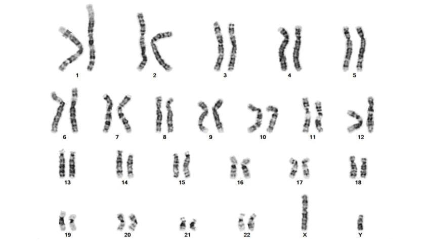
High-Resolution Brightfield and Fluorescence Imaging
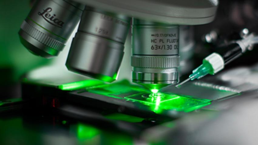
Built around Leica DM6 B microscope technology and a 12 MP CMOS camera, CytoVision DX provides automated brightfield and fluorescence imaging designed to capture chromosome banding patterns and fluorescence signals with exceptional clarity. Leica Biosystems states that the combination of high sensitivity and low-noise imaging helps support comprehensive chromosome review and detailed image analysis.
Flexible Capacity Designed for Laboratory Growth
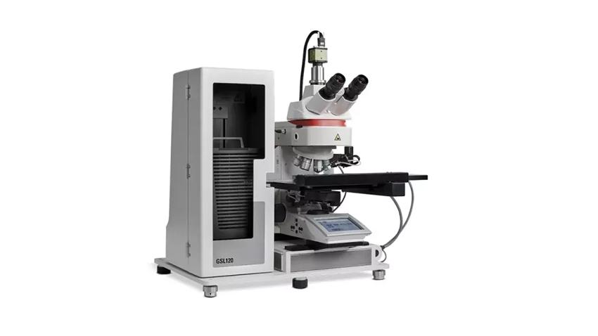
CytoVision DX is available in GSL 10 and GSL 120 configurations, providing batch capacities of 10 and 120 slides respectively. Hot-swappable slide trays help maintain productivity during scanning operations, while the system's compact footprint allows laboratories to maximize available workspace. Together, these capabilities help laboratories scale operations while maintaining workflow efficiency.
Connect Teams, Sites, and Laboratory Systems
Designed for modern laboratory environments, CytoVision DX supports remote review workflows, director oversight, user access controls, and multi-site collaboration. The platform also supports interoperability with third-party laboratory systems to facilitate data management, workflow integration, and collaboration across distributed teams and locations.
Built on Decades of Cytogenetics and Imaging Experience
CytoVision DX builds on more than 30 years of CytoVision experience and incorporates Leica Biosystems imaging expertise. The platform is compatible with Windows 11 and includes user administration capabilities for modern IT environments. Combined with Leica Biosystems' long-standing presence in pathology, microscopy, and regulated healthcare technologies, CytoVision DX provides laboratories with a solution backed by extensive scientific and engineering experience.
Specifications
Humidity: 20-90% non-condensing (38.7°C)
Ambient Temperature: 15° to 35°C (59° to 95°F)
Humidity: 20-70% non-condensing Maximum relative humidity: 70% for temperatures up to 36ºC (96.8ºF)
(included with all scanning systems)
(included with fluorescence scanning systems)
CytoVision DX Product Configuration Comparison
| Configuration |
Review Station |
Capture Station without Microscope |
Capture Station with Microscope |
Scanning Station with GSL 10 Automated Slide Loader |
Scanning Station with GSL 120 Automated Slide Loader |
|---|---|---|---|---|---|
| CytoVision DX software application & software protection key |
|
|
|
|
|
| Workstation – Computer with Windows OS, Display Monitor, and Uninterrupted Power Supply (can be provided by Leica Biosystems or end user) |
|
|
|
|
|
| 12 megapixel CMOS camera |
|
|
|
|
|
| Xylis LED lamp for FISH station |
|
Optional |
|
|
|
| Leica BM6 B automated microscope |
|
|
|
|
|
| XY stage |
|
|
Mechanical | Motorized | Motorized |
| Automated oiler |
|
|
|
|
|
| Barcode reader |
|
|
|
|
|
| Automated Slide loader capacity |
|
|
|
10 slides | 120 slides |
Downloads
Knowledge Hub
CytoVision DX is an image capture, analysis, and case-management system designed for clinical cytogenetics laboratories. It supports automated brightfield and fluorescence imaging as part of a workflow from slide capture to case review and reporting.
Karyotyping, interphase FISH, tissue FISH scanning, spot-counting capture, and m-FISH analysis are supported applications.
Yes. CytoVision DX's mixed-batch scanning supports karyotyping, interphase FISH, and tissue FISH imaging in a single session.
The system features a Leica DM6 B microscope and a 12 MP CMOS camera for brightfield and fluorescence imaging. Included objectives depend on the scanning configuration; options include 1.25x, 10x, 20x, and 100x oil objectives, with a 63x oil objective, fluorescence light source, and filters included with fluorescence scanning systems.
The system automates the identification, location, and high-resolution capture of interphase and metaphase cells. This reduces hands-on processes and allows technologists to focus on analysis.
Yes. Remote review, user-access controls, and multi-site collaboration are among the system’s connectivity capabilities.
The difference is batch capacity: 10 slides for GSL 10 and 120 slides for GSL 120. Laboratories should also evaluate their application mix, daily and peak workload, automation needs, physical space, staffing model, and integration requirements with a Leica Biosystems representative.
Hardware installation, application-software installation, training, maintenance and troubleshooting, cybersecurity, and safety are key implementation components. Exact responsibilities, timelines, validation activities, and infrastructure requirements should be confirmed for the selected configuration and local laboratory environment.